
Fig. 10
- ID
- ZDB-IMAGE-170621-16
- Publication
- Solek et al., 2017 - Lineage tracing of dlx1a/2a and dlx5a/6a expressing cells in the developing zebrafish brain
- All Figures
- Figures for Solek et al., 2017
|
Fig. 10
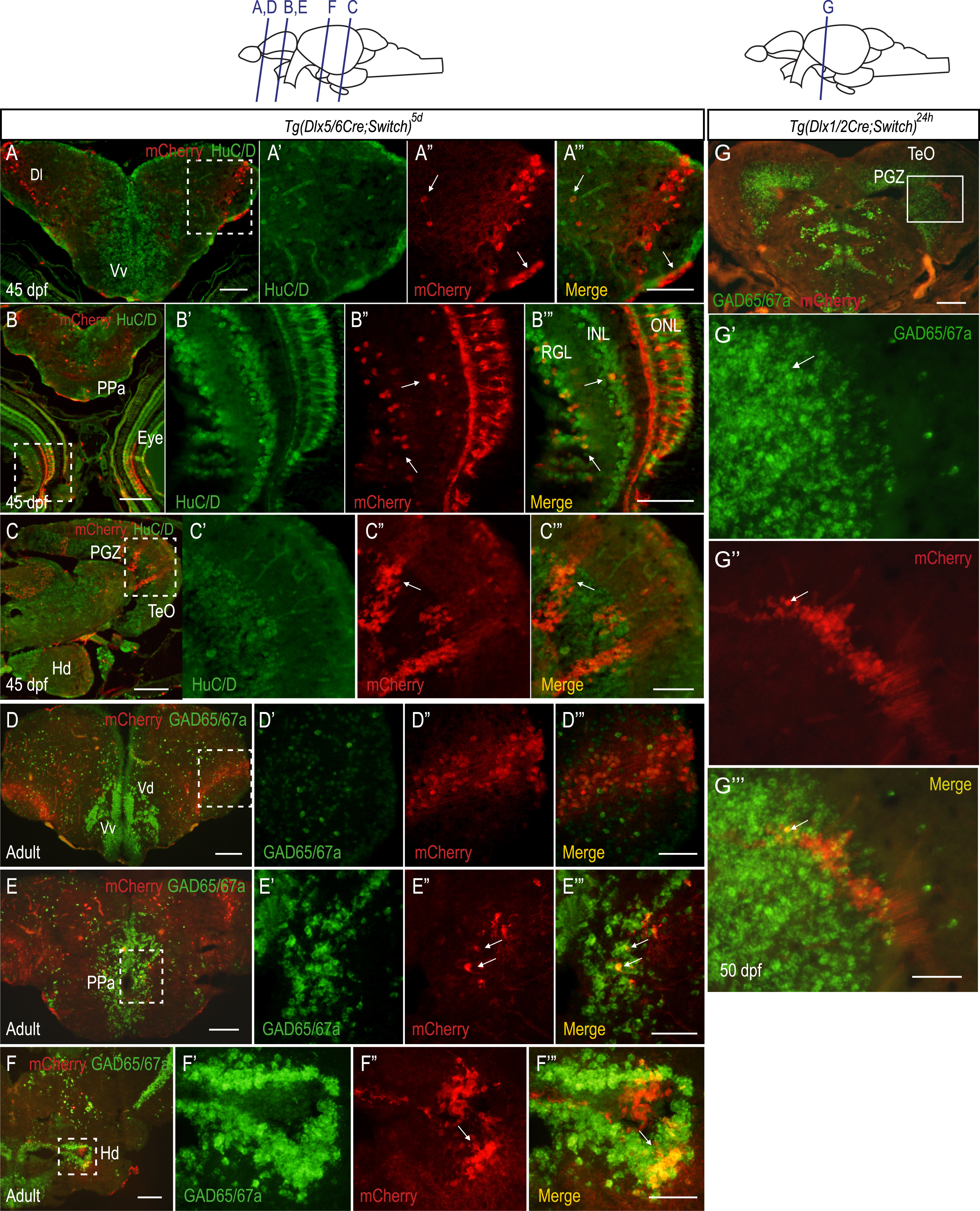
Most of the supernumerary mCherry expressing cells in juvenile and adult Tg(dlx5/6Cre;Switch)5d brains do not express gad65/67a. (A-C) Double IHC of juvenile (45 dpf) brains for mCherry (red) and HuC/D (green). n=3 in two biological replicates. Examples of cells expressing mCherry and HuC/D in the telencephalic area, the retina and the PGZ are indicated by white arrows in A''-C'' and A'''-C'''. (D-F) ISH for gad65 and gad67a probes (green) coupled with immunohistochemistry against mCherry proteins (red) on cryosections of adult (>6 months post-fertilization) brains. Examples of cells expressing mCherry and gad65/67a in the PPa near the ventricular zone (E) and in the Hd (F) are indicated by white arrows in E'', E''', F'' and F'''. n=4 in two biological replicates. (G) ISH for gad65 and gad67a probes (green) coupled with immunohistochemistry against mCherry proteins (red) on cryosections of Tg(dlx1/2Cre;Switch)24h juvenile brain showing modest expansion of mCherry cells in the PGZ. An example of a single cell that expresses both mCherry and gad65/67a is indicated by a white arrow in G', G'' and G'''. Scale bar: A-E: 50 µm; A'''-E''': 20 µm.
Reprinted from Developmental Biology, 427(1), Solek, C.M., Feng, S., Perin, S., Weinschutz Mendes, H.C., Ekker, M., Lineage tracing of dlx1a/2a and dlx5a/6a expressing cells in the developing zebrafish brain, 131-147, Copyright (2017) with permission from Elsevier. Full text @ Dev. Biol.