
Fig. 5 S1
- ID
- ZDB-IMAGE-170615-2
- Publication
- Hockman et al., 2017 - Evolution of the hypoxia-sensitive cells involved in amniote respiratory reflexes
- All Figures
- Figures for Hockman et al., 2017
|
Fig. 5 S1
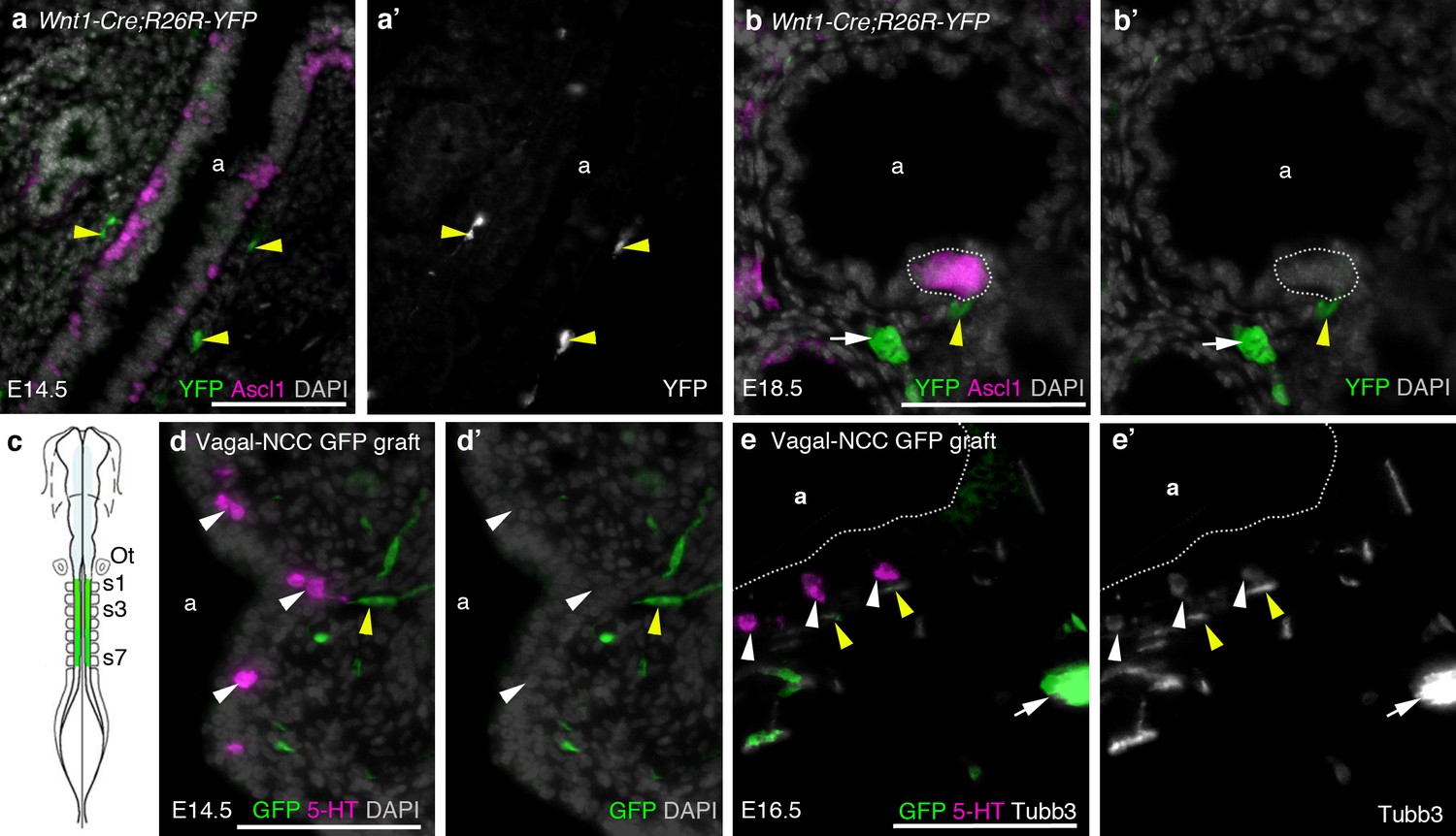
The neural crest does not contribute to amniote PNECs.
(a–b') Transverse sections through the lungs of Wnt1-Cre;R26R-YFP mouse embryos, in which neural crest cells are permanently labeled with YFP (Danielian et al., 1998; Srinivas et al., 2001), at E14.5 (a,a') and E18.5 (b,b'). PNECs (Ascl1 [Mash1]-positive cells in the airway epithelium; Ito et al., 2000) are unlabeled, whether solitary or clustered (white dotted line in b,b'), although nearby neural crest-derived cells in the subjacent mesenchyme (yellow arrowheads) are YFP-positive, including putative Schwann cells on a nerve innervating the PNECs (yellow arrowheads in b,b') and an intrinsic pulmonary ganglion (white arrow in b,b'), as expected (Freem et al., 2010). (In a,a’, the two fainter, out-of-focus green spots within the epithelium are background artefacts from the anti-GFP immunostaining.) (c) The vagal neural crest was labeled in the chicken using GFP-transgenic to wild-type neural tube grafts at E1.5 (schematic modified from Le Douarin, 2004). (d–e') Transverse sections through the lungs of grafted embryos at E14.5 (d,d') and E16.5 (e,e'). PNECs (serotonergic cells in the lung airway epithelium) are unlabeled (white arrowheads), while putative Schwann cells (elongated cells, yellow arrowheads) and a nearby intrinsic pulmonary ganglion (white arrow) are GFP-positive, as expected (Burns and Delalande, 2005). 5-HT, serotonin; a, airway; Ot, otic vesicle; s, somite. Scale-bars: 10 μm in a; 50 μm in b,d,e.