
Fig. 1
- ID
- ZDB-IMAGE-170614-24
- Genes
- Antibodies
- Publication
- Hockman et al., 2017 - Evolution of the hypoxia-sensitive cells involved in amniote respiratory reflexes
- All Figures
- Figures for Hockman et al., 2017
|
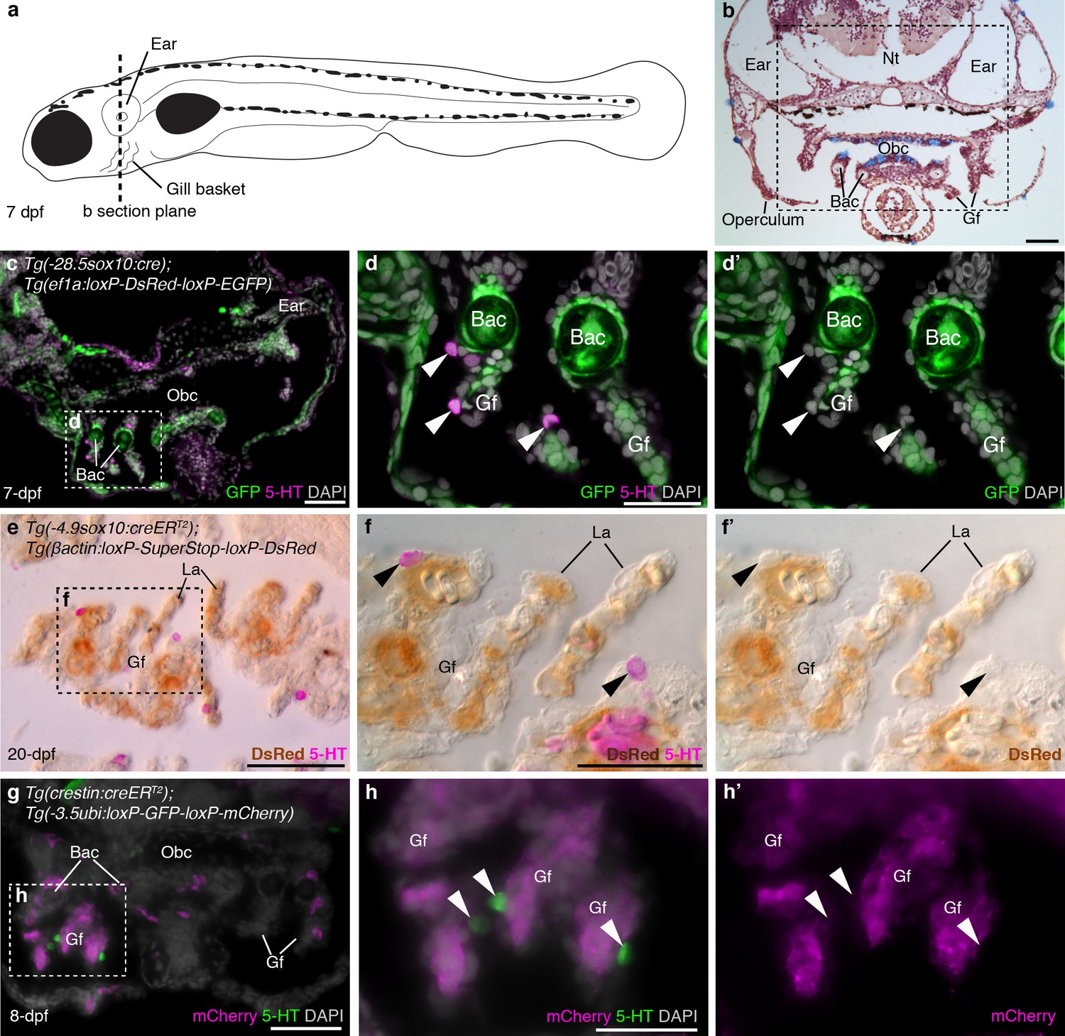
Fig. 1
Zebrafish NECs are not neural crest-derived: genetic lineage-tracing data.
(a) Schematic of a 7–8 dpf zebrafish; dotted line indicates section plane in b-f'. (b) Hematoxylin and eosin staining at 7-dpf reveals gill filaments branching from branchial arch cartilages, and the orobranchial cavity. Dashed box indicates approximate region in c,g. (c–d') In 7-dpf Tg(-28.5sox10:cre);Tg(ef1a:loxP-DsRed-loxP-EGFP) zebrafish, GFP labels neural crest-derived branchial arch cartilage and mesenchyme, but not NECs in the gill filaments (identified by immunoreactivity for serotonin, 5-HT; arrowheads). (e–f') Horizontal section through the gills of a 20-dpf Tg(-4.9sox10:creERT2);Tg(βactin:loxP-SuperStop-loxP-DsRed) zebrafish. DsRed (brown precipitate) labels neural crest-derived gill pillar cells, but not NECs (arrowheads; inverted fluorescent image overlaid on bright-field image). (g–h') In 8-dpf Tg(crestin:creERT2);Tg(-3.5ubi:loxP-GFP-loxP-mCherry) zebrafish, mCherry labels gill pillar cells but not NECs (arrowheads). 5-HT, serotonin; Bac, branchial arch cartilage; Gf, gill filament; La, lamellae; Nt, neural tube; Obc, orobranchial cavity. Scale-bars: 50 μm in b,c,e,g; 25 μm in d,f,h.