
|
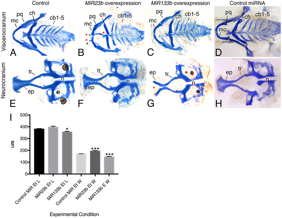
Fig. 7
Figure 7. Injection of MiR133b and MiR23b RNA duplexes resulted in facial defects. Alcian blue stain of 6 dpf zebrafish larvae, flat mounted ventral views, anterior is to the left. (A,D,E,H) Control embryos, either uninjected or 33 μM control miR-injected, display wild type type craniofacial cartilage in both the viscerocranium and neurocranium. (B,F) Injection of 33 μM RNA duplex of MiR23b leds to a slight increase in the width of the ethmoid plate, likely resulting from shortening of the trabeculae (tr) (F), and production of ectopic cartilage (*; B) in the B) in the viscerocranium. (C,G) Injection of 6.25 μM RNA duplex of MiR133b resulted in hypoplastic cartilage structure and a cleft (*) in the ethmoid plate (ep; G,I). (I) Quantification of ethmoid plate length and width comparing standard control MiR (n = 10) injected with 33 μM compared to MiR23b (n = 9) and MiR133b (n = 9). Elements were measured and the mean and standard deviation for each are: Ethmoid plate length for control MiR, 380 ± 13.23 μm; MiR23b, 396 ± 27.13 μm; and MiR133b, 355 ± 19.27 μm; Ethmoid plate width for control MiR, 169.2 ± 4.92 μm; MiR23b, 193.9 ± 193.9 ± 14.09 μm; and MiR133b: 144.7 ± 14.7μm. Student's (Welsh's) T-test was used to compare to the standard control using GraphPad PRISM. *p < 0.02 and ***p < 0.0008. Anterior is to the left. cb, ceratobranchial; ch, ceratohyal; mc, Meckel's cartilage; n, notochord; pq, palatoquadrate.