
Fig. 5 S1
- ID
- ZDB-IMAGE-170609-22
- Publication
- Venero Galanternik et al., 2017 - A novel perivascular cell population in the zebrafish brain
- All Figures
- Figures for Venero Galanternik et al., 2017
|
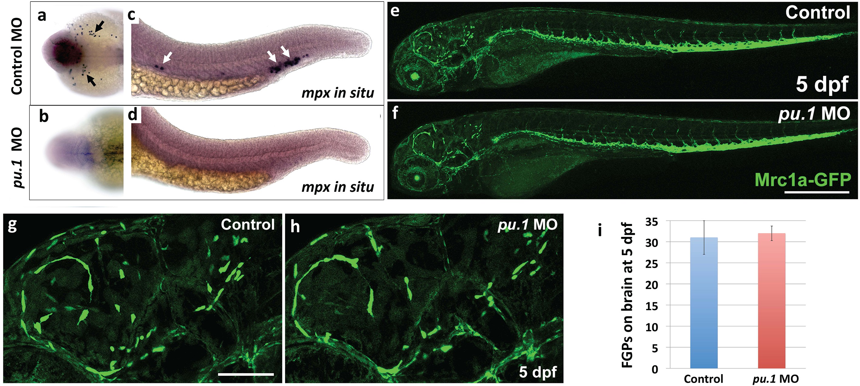
Fig. 5 S1
FGPs are not derived from primitive hematopoiesis.
(a-d) Dorsal yolk (a,b) and lateral trunk (c, d) transmitted light images of 20 hpf control morpholino (a,c) or pu.1 morpholino (b,d) injected embryos subjected to whole mount in situ hybridization and probed for mpx. Arrows in panels a and c show mpx-positive cells in controls that are absent in pu.1 morpholino-injected animals.( e,f) Lateral view confocal images of 5 dpf Tg(mrc1a:eGFP) transgenic control morpholino (e,g) or pu.1 morpholino (f, h) injected larvae. (g,h) Higher magnification images of brains of the same control morpholino (g) or pu.1 morpholino (h) injected larvae shown in panels e and f, respectively. (i) Quantification of FGP cells present in control vs. pu.1 morpholino-injected animals at 5 dpf (t-test, p-value=0.76). Scale bars: 500 µm (e,f), 200 µm (g,h).