
Fig. 4 S2
- ID
- ZDB-IMAGE-170609-20
- Publication
- Venero Galanternik et al., 2017 - A novel perivascular cell population in the zebrafish brain
- All Figures
- Figures for Venero Galanternik et al., 2017
|
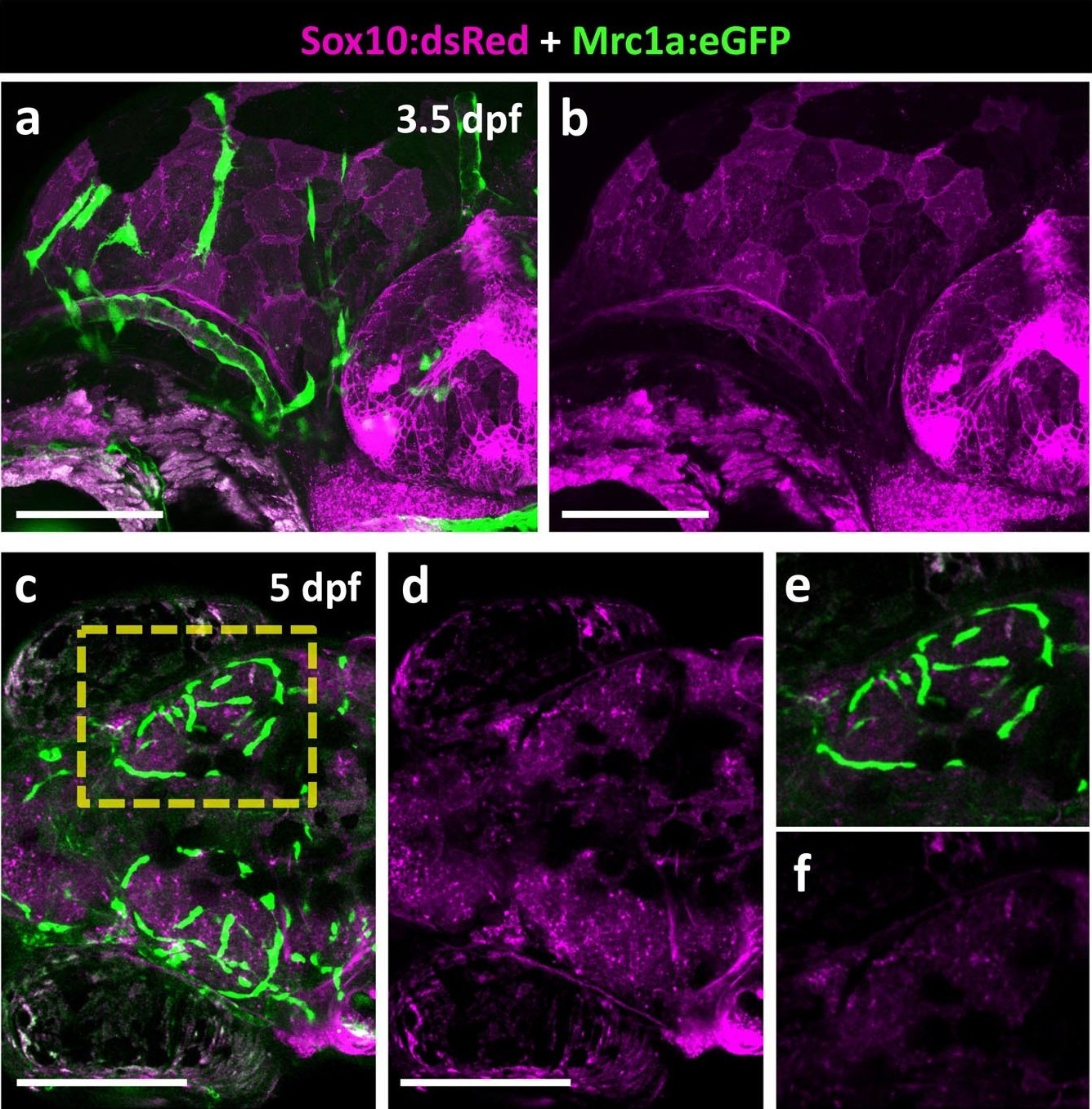
Fig. 4 S2
FGPs are not Neural Crest derived.
(a-b) Lateral view of a 3.5 dpf Tg(sox10:dsRed); Tg(mrc1a:eGFP) double transgenic embryo showing mrc1a:eGFP-positive FGPs in green (a) and Sox10:dsRed–positive neural crest derived structures in magenta (a,b; n = 5 animals imaged). (c–f), Dorsal views of the optic lobes of a 5 dpf Tg(sox10:dsRed); Tg(mrc1a:eGFP) double transgenic embryo with mrc1a:eGFP-positive FGPs in green (c,e) and Sox10:dsRed–positive neural crest derived structures in magenta (c-f; n = 5 imaged animals). (e,f) Higher magnification images of the right optic lobe in the yellow box in panel c. Scale bars: 100 μm (a, b), 200 μm (c, d).