
|
Fig. 5
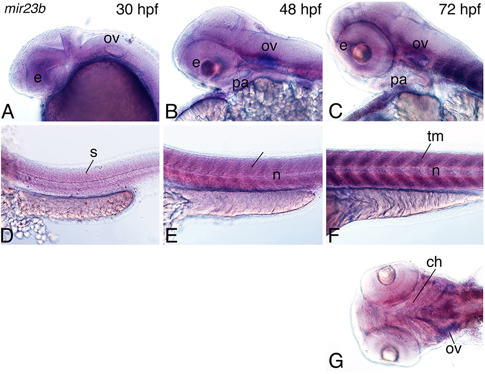
Figure 5. Expression of mir23b in zebrafish embryos. Whole-mount in situ hybridization analysis with a digoxigenin-labeled probe against the mir23b transcript at 30–72 hpf. (A–C) Expression is observed in the otic vesicle (ov; A), weakly in the eye (e), and pharyngeal arch (B,C), and palate (pl; C). (D–F) In the trunk, expression is observed in the trunk muscle the somites (s; E,D) at 30, 48 hpf, and trunk muscle (tm; F) at 72 hpf. (G) Ventral view of expression shows more detailed expression in the cranial muscle including the muscles supporting the craniofacial structures (arrows) and the eye. Anterior is to the left. n, notochord; ch, ceratohyal.