
IMAGE
Fig. 2
- ID
- ZDB-IMAGE-170607-2
- Genes
- Publication
- Umans et al., 2017 - CNS angiogenesis and barriergenesis occur simultaneously
- All Figures
- Figures for Umans et al., 2017
Image
|
Figure Caption
Fig. 2
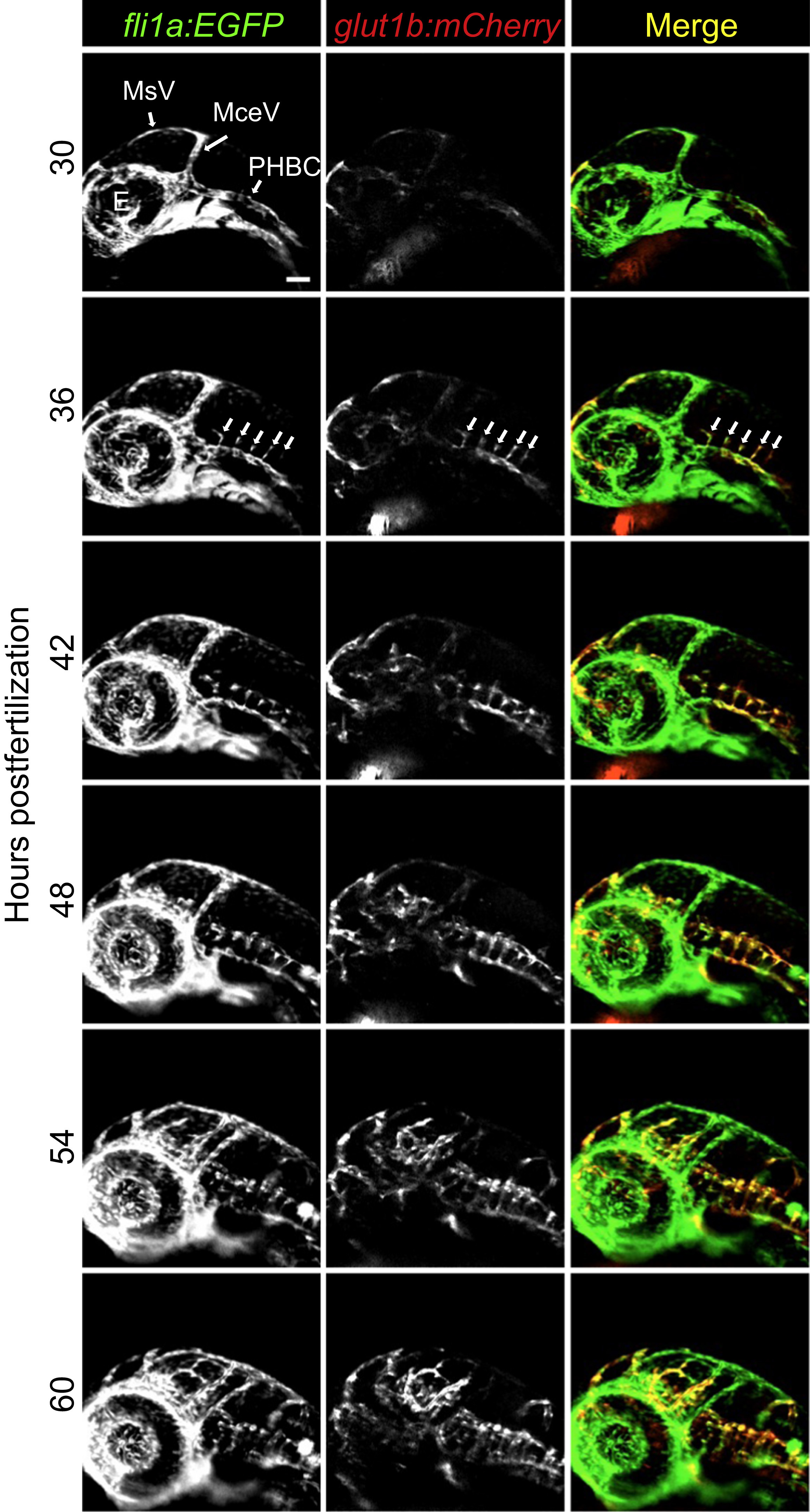
CNS angiogenesis and barriergenesis occur simultaneously. glut1b:mCherry; fli1a:EGFP double transgenic embryos were imaged by time-lapse confocal microscopy. Shown here are snapshots at 6-h intervals beginning at the onset of CNS angiogenesis (30 hpf). Note that mCherry is immediately expressed as brain endothelial cells migrate into the brain parenchyma from the PHBCs (arrows in 36 hpf panels). See 30 h time-lapse Videos 1 for more detail. MsV, mesencephalic vein; MCeV, mid-cerebral vein; PHBC, primordial hindbrain channel, E, eye. Scale bar in top left image is 50 µm for all images.
Figure Data
Acknowledgments
This image is the copyrighted work of the attributed author or publisher, and
ZFIN has permission only to display this image to its users.
Additional permissions should be obtained from the applicable author or publisher of the image.
Reprinted from Developmental Biology, 425(2), Umans, R.A., Henson, H.E., Mu, F., Parupalli, C., Ju, B., Peters, J.L., Lanham, K.A., Plavicki, J.S., Taylor, M.R., CNS angiogenesis and barriergenesis occur simultaneously, 101-108, Copyright (2017) with permission from Elsevier. Full text @ Dev. Biol.