
Image
|
Figure Caption
Fig. S8
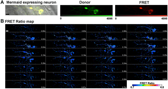
Mosaic expression of HuC:Mermaid vector in a 20 hpf zebrafish spinal motor neuron, and a FRET ratio map of a spontaneous depolarisation event.
(A) In order to verify whether the aberrant motor phenotype observed in 20 hpf mSod1 embryos was associated with alterations in the spontaneous depolarisation of spinal neurons, one-cell-stage embryos were micro-injected with the FRET-based voltage biosensor Mermaid ORF under the control of the pan-neuronal promoter HuC. The bright field image merged with the fluorescence signal shows efficient biosensor expression in a spinal motor neuron. The detected donor and FRET channel are also shown. Scale bar: 10 µm. (B) The time points on the FRET ratio map of the same motor neuron during a spontaneous depolarisation event shows that the FRET ratio increases with membrane potential. Scale bar: 10 µm.
Acknowledgments
This image is the copyrighted work of the attributed author or publisher, and
ZFIN has permission only to display this image to its users.
Additional permissions should be obtained from the applicable author or publisher of the image.
Full text @ Sci. Rep.