
Fig. 2
- ID
- ZDB-IMAGE-170607-11
- Genes
- Antibodies
- Publication
- Benedetti et al., 2016 - INaP selective inhibition reverts precocious inter- and motorneurons hyperexcitability in the Sod1-G93R zebrafish ALS model
- All Figures
- Figures for Benedetti et al., 2016
|
Fig. 2
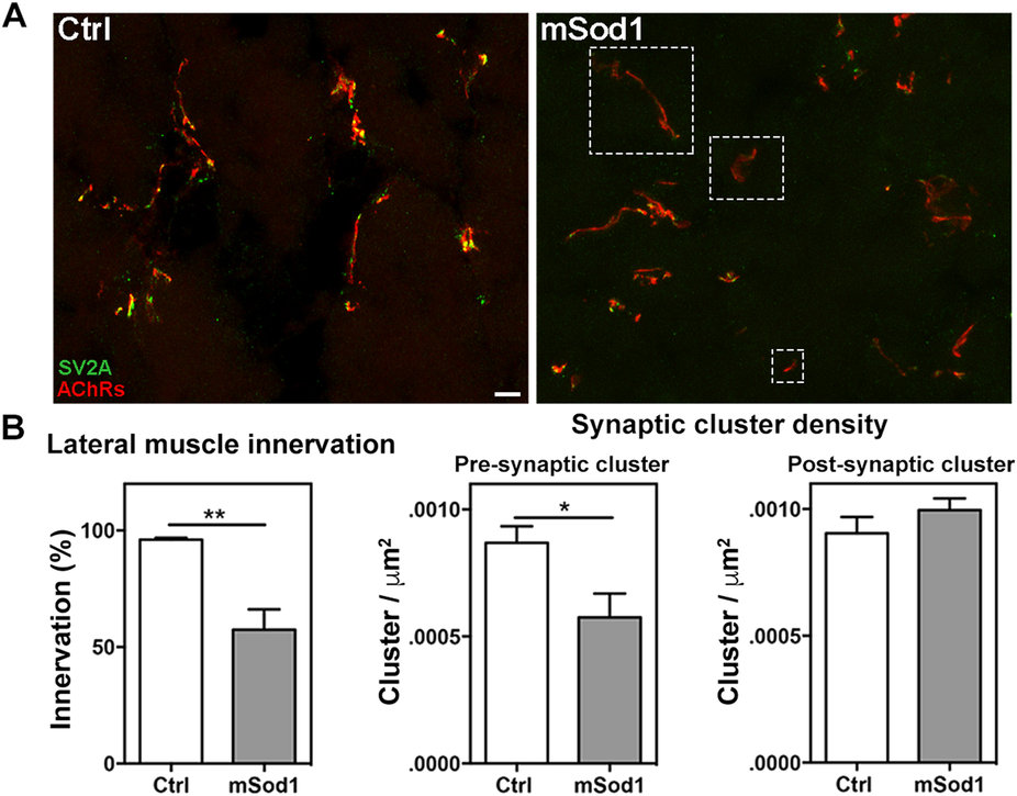
Twelve-month-old mSod1 zebrafish show compromised lateral white muscle innervation.
(A) Maximum projections of confocal images of synaptic vesicle protein 2A (SV2A, green) and muscle acetylcholine receptors (AChRs, red) covering the entire thickness (20 μm) of a Ctrl and mSod1 zebrafish lateral muscle cryostat section. In the Ctrl zebrafish, each post-synaptic specialization enriched with acetylcholine receptors faces motor nerve terminals containing vesicle clusters, whereas many of the post-synaptic clusters in the mSod1 section lack an association with motor pre-synaptic terminals (white dashed boxes). Scale bar: 20 μm. (B) The percentage of innervation of post-synaptic specializations is significantly reduced in the mSod1 zebrafish (57.48 ± 8.69% vs 95.94 ± 0.84%), and three-dimensional co-localization analysis of z-stacks covering the entire thickness of the sections revealed a significant reduction in pre-synaptic cluster density (5.76 ± 0.09 vs 8.69 ± 0.06 × 10⁻⁴ clusters/μm2) but not in post-synaptic cluster density (9.96 ± 0.05 vs 9.04 ± 0.06 × 10⁻⁴ clusters/μm2). The columns in each graph indicate the mean value ± SEM of the indicated parameter in five Ctrl and six mSod1 zebrafish. The measures were statistically analyzed using an unpaired Student t-test (*P < 0.05; **P < 0.01).