
|
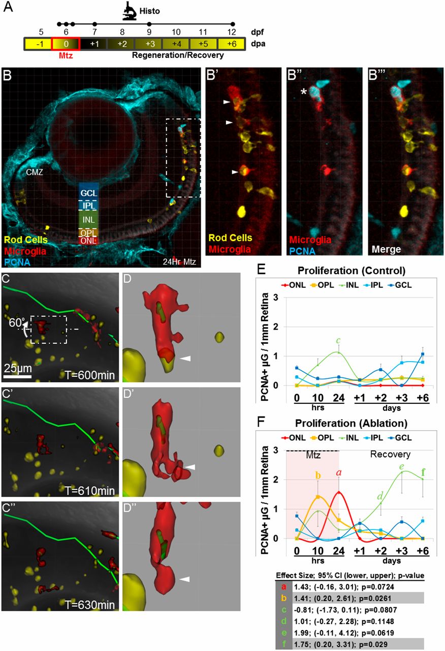
Fig. S3
Microglia exhibit additional hallmarks of innate immune cell activation following rod cell ablation. (A) Schematic of assay timelines for data shown in B–F. (B) Representative retinal section of 7-dpf rho:YFP-NTR;mpeg1:Tomato larvae immunostained for PCNA (blue) after 24-h exposure to Mtz (56× magnification). The boxed region in B (enlarged in B′ and B′′) shows evidence of microglia phagocytosis of rod cell debris and microglia proliferation. Arrowheads in B′ denote yellow rod cell debris within red microglia. The asterisk in B′′ denotes red/blue double-positive microglia. A merged image is shown in B′′′. (C–C′′) Time-lapse sequence showing microglia (red cell) phagocytosis of rod cell debris (yellow). (D–D′′) The boxed region in C was enlarged and rotated 60° to provide additional detail (arrowheads). (E and F) Quantification of microglia proliferation (PCNA immunoreactivity) within defined somal regions in untreated control (E) and Mtz-treated (F) retinas, plotted over time. (E) Control retinas showed little to no proliferation in most somal layers. (F) Mtz-treated retinas showed increased microglia proliferation in photoreceptor regions (OPL, yellow line; ONL, red line) during the time of rod cell death and in the INL (green line) at later time points. Paired statistical comparisons (Student’s t test) made between time point-matched control (E) and experimental (F) data are denoted by a unique letter color coded according to retinal layer. Error bars in E and F indicate SEM. Effect size, 95% confidence intervals, and P values are provided in the table. (Note: Only pairs showing statistically validated differences are listed.) CMZ, ciliary margin zone.