
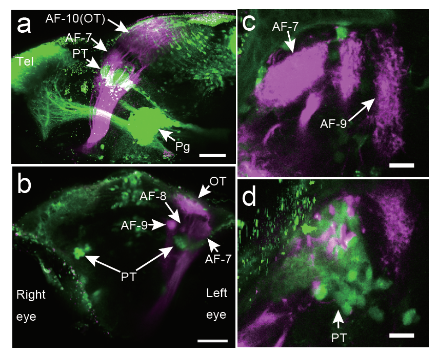
Fig. S13
- ID
- ZDB-IMAGE-170526-10
- Publication
- Muto et al., 2017 - Activation of the hypothalamic feeding centre upon visual prey detection
- All Figures
- Figures for Muto et al., 2017
|
Fig. S13
Locations of the 119B-pretectal area and the retinal ganglion cell arbours
Locations of the axonal arbourisation fields (AFs) of the ganglion cells (magenta) relative to the gSAzGFFM119B-labelled cells (green). DiI was injected into the right eye, and the left eye was removed for observation. a, Lateral view from the left side. Scale bar: 50 μm. b, Front view. Scale bar: 50 μm. c, Top view (focused on the AF-7 area). Scale bar: 5 μm. d, Top view (focused 26 μm below the AF-7 area). Scale bar: 5 μm. PT, pretectal area; OT, optic tectum; Tel, telencephalon; Pg, preglomerular nuclei; D, dorsal; V, ventral; A, anterior; P, posterior.