
Fig. 3
- ID
- ZDB-IMAGE-170524-24
- Genes
- Publication
- Ibarra et al., 2017 - Dynamic regulation of Pin1 expression and function during zebrafish development
- All Figures
- Figures for Ibarra et al., 2017
|
Fig. 3
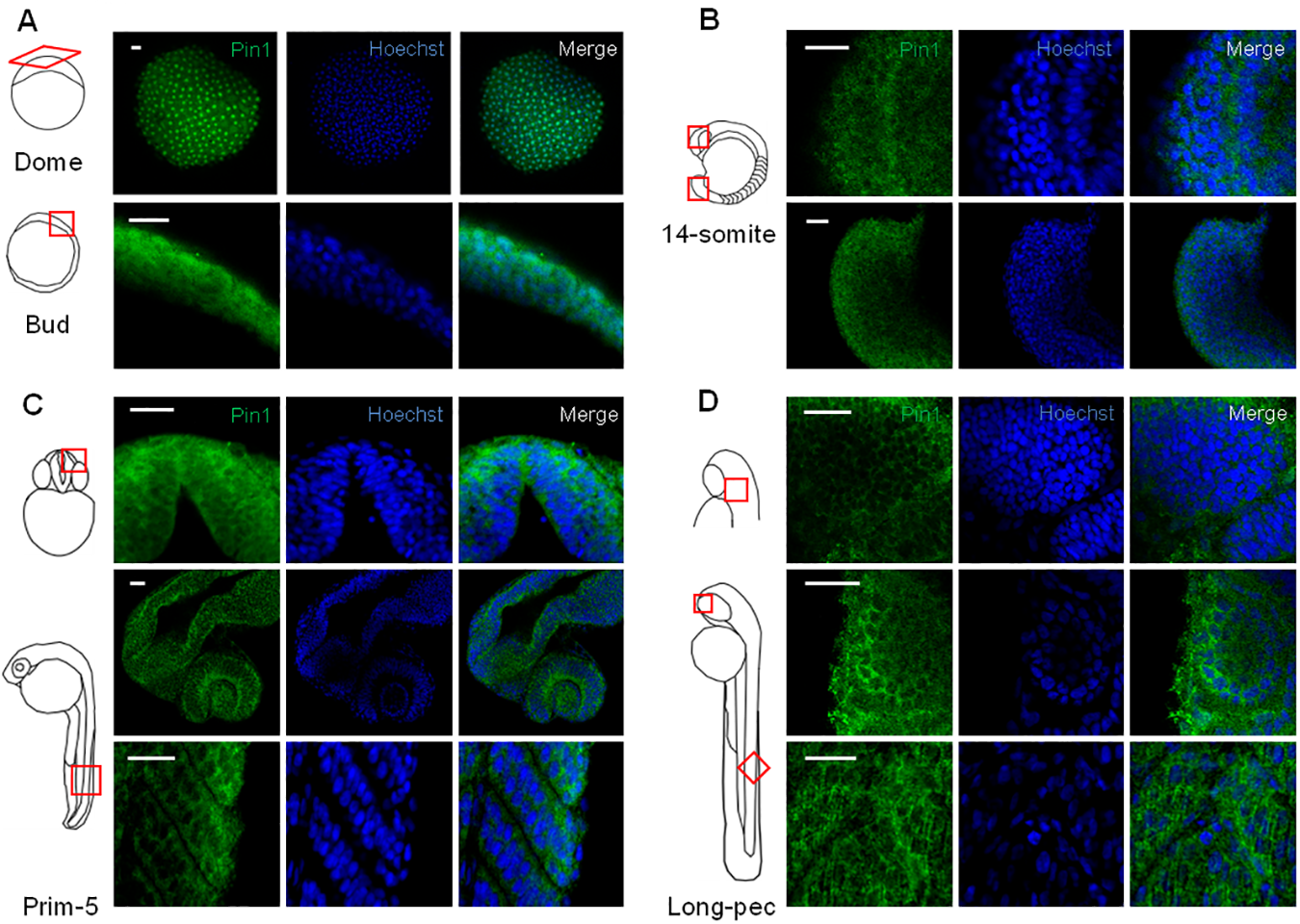
Analysis of Pin1 distribution in zebrafish embryos.
Confocal images of whole-mount immunofluorescence performed on embryos from different stages using Pin1 antibody. (A) Dome (4:30 hpf) and Bud stage (10 hpf). (B) 14-somite stage (16 hpf), lateral views from the head (upper panels) and tail (lower panels). (C) Prim-5 stage (24 hpf), frontal view of the head (upper panels), dorsolateral view of the head (middle panels) and lateral view of the tail. (D) Long-pec stage (48 hpf), dorsolateral view of the head (upper panels), lateral view of the head (middle panels) and lateral view of the tail (lower panels). Nuclei were stained with Hoechst. Scale bar = 25 μm.