
Image
|
Figure Caption
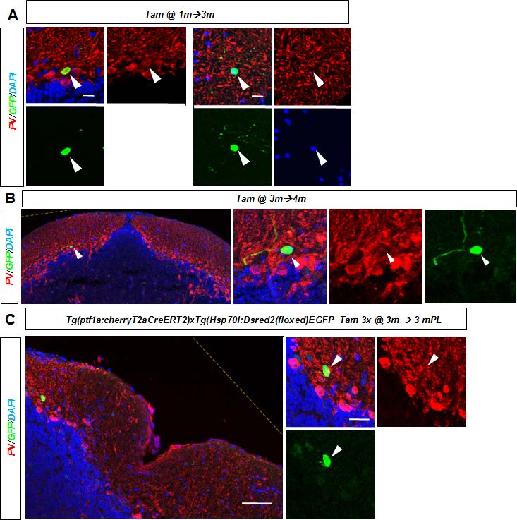
Fig. S4
A. Recombined GFP/PV+ Purkinje cell (left) and PV- negative inter-neuron (right) with stellate morphology in the cerebellum 3 months after recombination of a one month old juvenile fish. B. A recombined GFP/PV- inter-neuron in the adult cerebellum one month after tamoxifen treatment. C. A recombined GFP+ and PV- interneuron in the cerebellum three months after injury and tamoxifen treatment of an adult zebrafish. Yellow hatched line show original picture border in rotated in images.
Acknowledgments
This image is the copyrighted work of the attributed author or publisher, and
ZFIN has permission only to display this image to its users.
Additional permissions should be obtained from the applicable author or publisher of the image.
Full text @ Development