
|
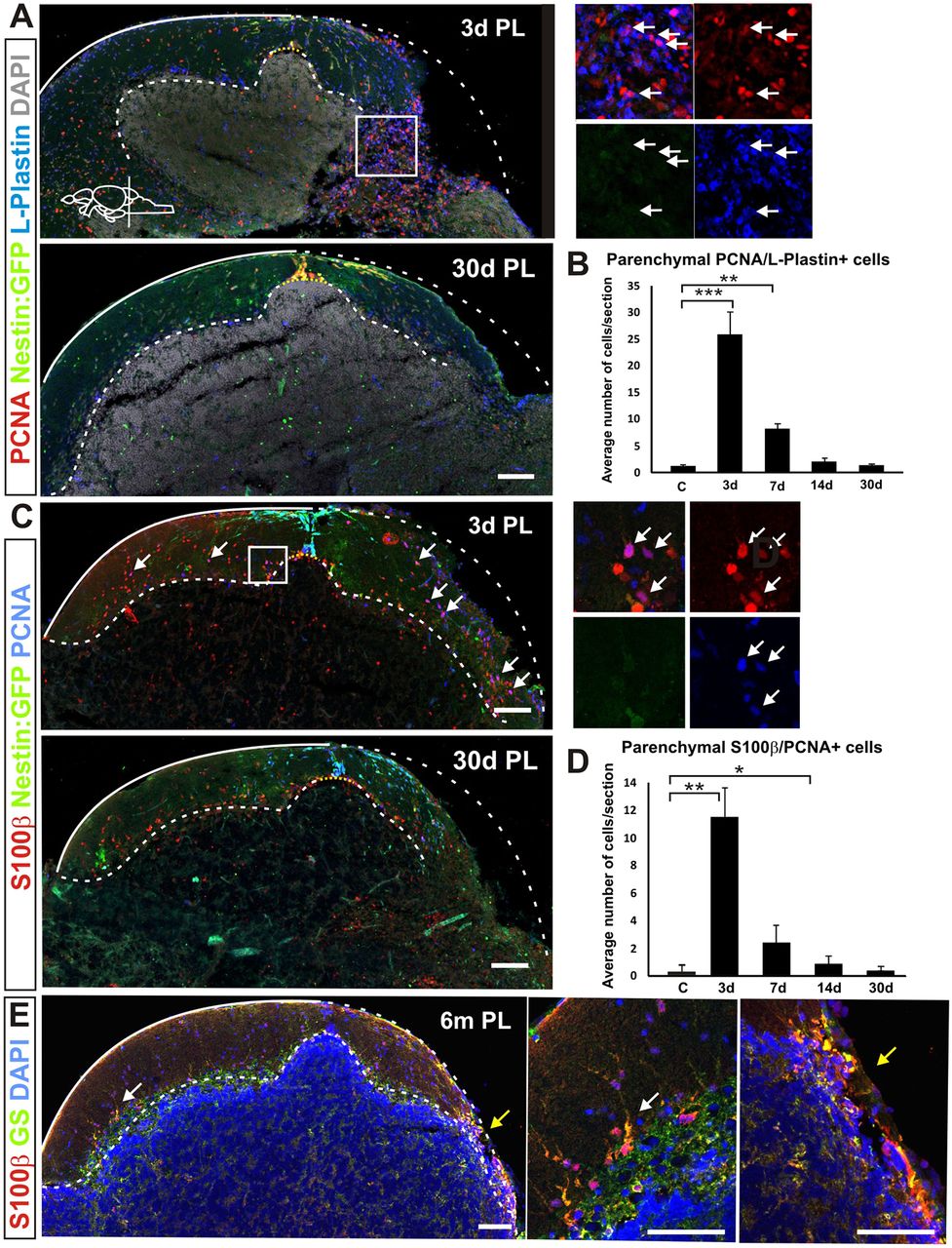
Fig. 3
Inflammation and scarring after injury. (A) Cerebellar transverse section showing proliferating (Pcna+) nestin:gfp− parenchymal cells (green) colocalising with the leukocyte marker L-plastin (arrows) 3 days after injury. Parenchymal proliferation and leukocyte numbers were restored towards homeostatic levels 30 days after injury. (B) Quantification of proliferating Pcna/L-plastin-positive cells in the cerebellar parenchyma after injury. A significant increase in the number of proliferating L-plastin+ cells is detected during the first week. The number of proliferating L-plastin+ cells is back to control levels 14 days after injury. **P<0.01, ***P<0.001; n=5 for each time point. (C) Proliferating Bergmann glia (Pcna/S100β+) are also detected in the parenchyma after injury (arrows). (D) Quantification of proliferating Bergmann glia shows a significant number in the cerebellar parenchyma 3 days after injury. *P<0.05, **P<0.01; n=55 for each time point. The number of proliferating Bergmann glia decreases the first week after injury and is back to homeostatic levels 30 days after injury. (E) Glial proliferation does not result in glial scarring. No notable accumulation of the glial markers S100β (red) and GS (green) can be detected at the injury site 6 months after injury (n=5). The arrowed regions are magnified to the right, illustrating proliferating leukocytes in A and proliferating Bergmann glia in C (white arrows) and Bergmann glia at the lesion site (yellow arrows). Scale bars: 100 μm.