
|
Fig. 1
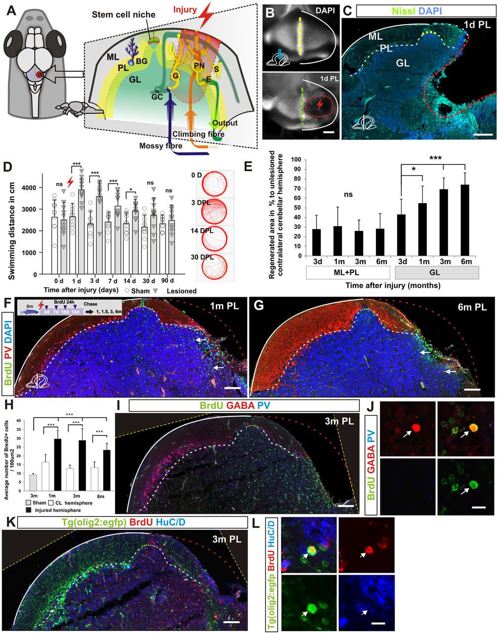
Morphological and behavioural recovery after cerebellar injury. (A) Overview of cerebellar architecture in zebrafish and the injury model. The cerebellum contains only a few cell types of distinct morphology. The cerebellum has a simple laminar architecture consisting of a molecular layer (ML), Purkinje cell layer (PL) and granule cell layer (GL). The unilateral injury removes cells from all three layers. Importantly, the lesion spares the stem cell niche. The GL consists of densely packed excitatory granule cells (GC) and inhibitory Golgi neurons (G). The PL contains Purkinje neurons (PN), specialised Bergmann glia (BG), and excitatory eurydendroid cells (E). The ML consists of nerve fibres and scattered inhibitory stellate cells (S). Neural stem cells are maintained in the dorsomedial part of the cerebellum around a remnant of the IVth ventricle. (B) A piece of the corpus cerebelli, including all cell layers, is unilaterally removed (red lightning). A confocal maximum projection showing a dorsal view of an unlesioned (top) and a lesioned (bottom) cerebellum 1 day after the injury. The stem cell niche is indicated with a yellow dashed line. The extent of the lesion is indicated with a red dashed line. (C) A transverse section of the corpus cerebelli showing the lesion 1 day after injury. (D) Automatic tracking of swimming distance and pattern during 5 minutes. Cerebellar injury induced significantly increased swimming activity from 1 to 14 days after injury. n=11 for sham and injured groups; 1, 3, 7 days post-lesion (DPL) ***P<0.001, 14 DPL *P<0.05; error bars indicate s.d. (E) Morphological quantification of the lesioned area after injury. Lesioned area compared with lesion size measured 3 days after injury. n≥5 per time point; 1 month *P<0.05, 3 and 6 months ***P<0.001; ns, not significant; error bars indicate s.d. (F,G) Transverse section of the corpus cerebelli showing that BrdU-stained cells (green) are recruited and maintained (arrows) in the GL of the lesioned hemisphere 1 and 6 months after injury. Few BrdU-stained cells are detected in the ML and PL. Purkinje cells are stained with parvalbumin (PV; red). (H) Quantification of BrdU-positive cells in the GL in the injured and contralateral hemisphere 1 , 3 and 6 months after injury. n=5 per time point; ***P<0.001; error bars indicate s.d. Sham control animal is included as reference point for normal granule cell production rate (3 months after BrdU pulse). CL, contralateral uninjured hemisphere. (I,J) Cerebellar transverse section showing BrdU, GABA and PV staining 3 months after injury. Very few BrdU/GABA-positive cells are detected (arrow). (K,L) Cerebellar transverse section showing olig2:gfp, BrdU and HuC/D staining 3 months after injury. BrdU/olig2:gfp-positive cells are very rarely detected (arrow). Yellow dashed line indicates original picture border in rotated images. Scale bars: 200 μm (B); 150 μm (C); 100 μm (F,G,I,K); 10 μm (J,L).