
Fig. S2
- ID
- ZDB-IMAGE-170512-22
- Genes
- Antibodies
- Publication
- Berberoglu et al., 2017 - Satellite-like cells contribute to pax7-dependent skeletal muscle repair in adult zebrafish
- All Figures
- Figures for Berberoglu et al., 2017
|
Fig. S2
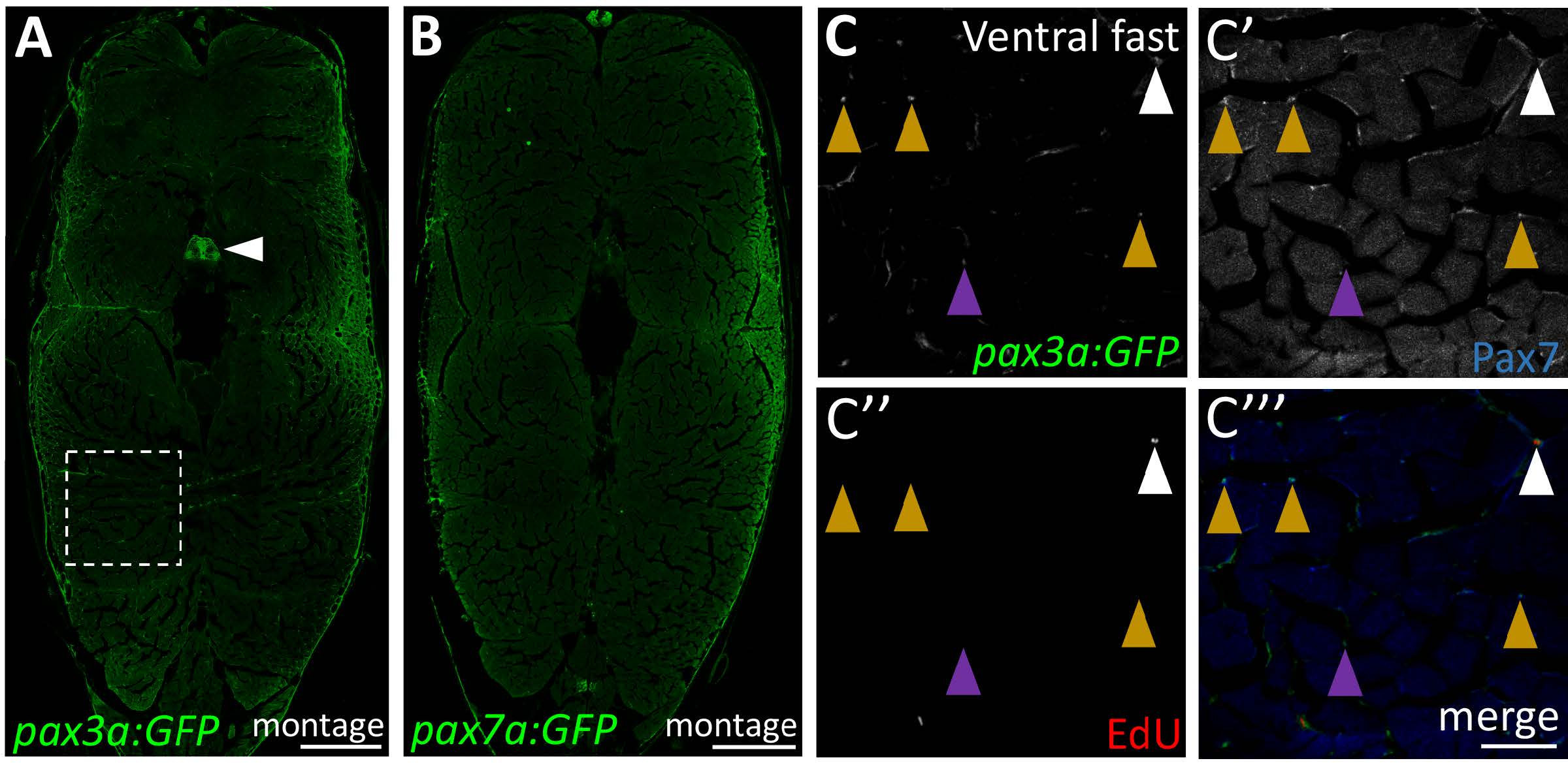
Characterization of pax3a:GFP transgenic expression within the adult zebrafish myotome. (A) Montage of entire cross-section showing pax3a:GFP transgene expression. In addition to satellite-like cells, pax3a:GFP is also expressed in the skin (outlining the section) and spinal cord (arrowhead). (B) Montage of entire cross-section showing pax7a:GFP transgene expression. In addition to satellite-like cells, pax7a:GFP is also expressed in the skin (outlining the section) and in a few cells within the spinal cord. (C-C''') Magnified view of ventral fast muscle region (approximate region of analysis is indicated by the box in A). Satellite-like cells expressing pax3a:GFP and Pax7 (mustard arrowheads indicate examples) are sparsely present within the ventral fast muscle region. Although the majority of pax3:GFP-expressing cells express Pax7, there are some pax3a:GFP-positive cells that are Pax7-negative (purple arrowhead) that may represent cells with GFP-positive cytoplasm for which the nucleus is on an adjacent section. Scattered EdU-positive cells are present throughout the adult myotome (white arrowhead). Scale bar in A and B is 300 μm and in C''' is 75 μm.
Reprinted from Developmental Biology, 424(2), Berberoglu, M.A., Gallagher, T.L., Morrow, Z.T., Talbot, J.C., Hromowyk, K.J., Tenente, I.M., Langenau, D.M., Amacher, S.L., Satellite-like cells contribute to pax7-dependent skeletal muscle repair in adult zebrafish, 162-180, Copyright (2017) with permission from Elsevier. Full text @ Dev. Biol.