
Fig. S3
- ID
- ZDB-IMAGE-170512-21
- Publication
- Berberoglu et al., 2017 - Satellite-like cells contribute to pax7-dependent skeletal muscle repair in adult zebrafish
- All Figures
- Figures for Berberoglu et al., 2017
|
Fig. S3
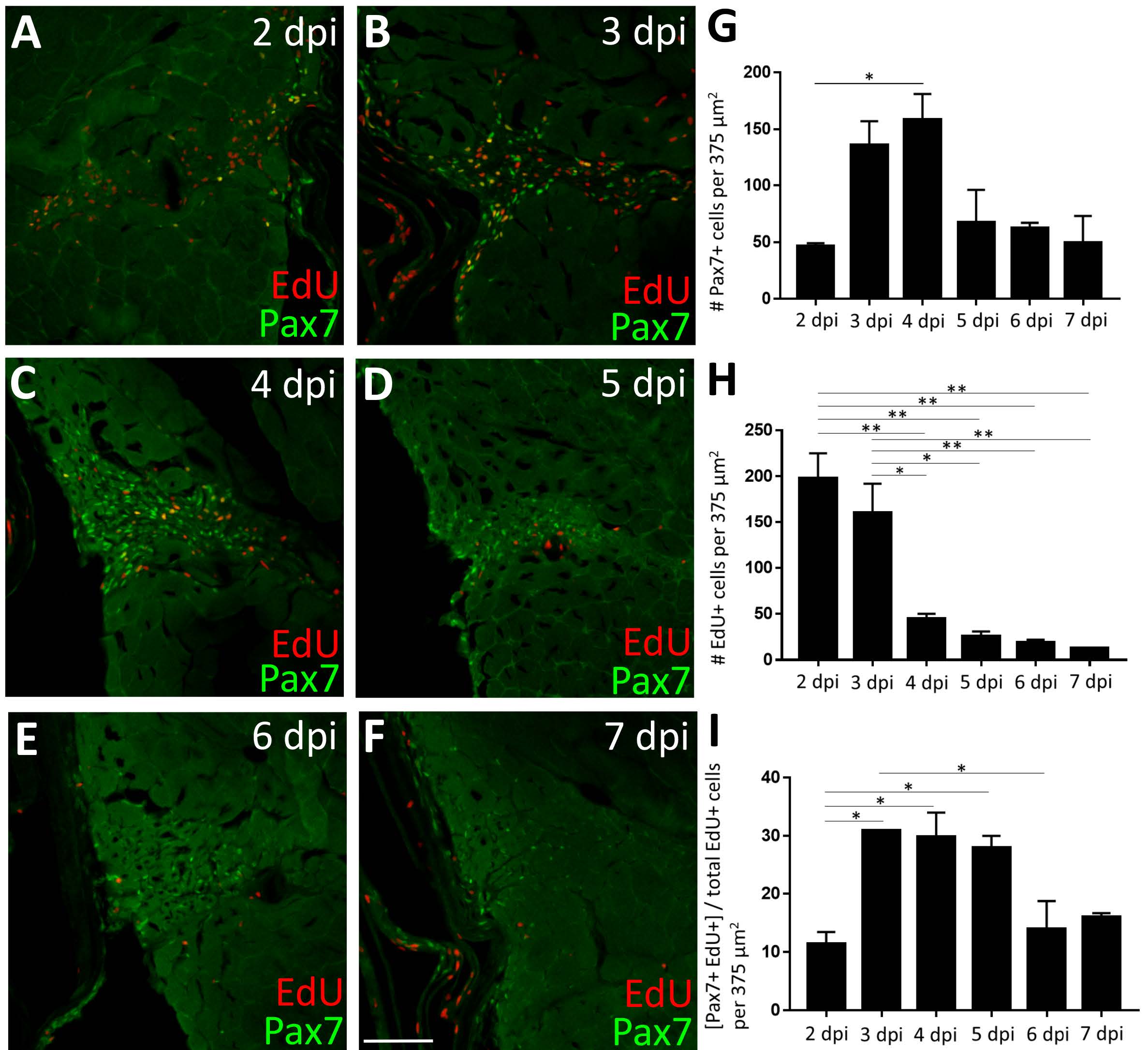
By 3 dpi, a significant number of proliferating cells in the injured myotome express Pax7. (A-F) Pax7 expression (green) at post-injury time-points 2-7 dpi, with proliferating cells labeled via an acute EdU pulse (red). The image in B is also shown in Fig. 3D. (G) Pax7-positive cell number increases significantly between 2 and 4 dpi. (H) EdUpositive cell number is highest at 2 and 3 dpi and declines thereafter. (I) When the number of Pax7-positive cells is at their peak (3-4 dpi, see G), they represent ~30% of the total EdUpositive cell population, indicating that additional non-satellite-like cells are also proliferating at the injury site. One-way ANOVA followed by Tukey’s multiple comparisons test was performed for all of the above statistical analyses; p*<0.05, p**<0.01. Scale bar in F is 75 μm.
Reprinted from Developmental Biology, 424(2), Berberoglu, M.A., Gallagher, T.L., Morrow, Z.T., Talbot, J.C., Hromowyk, K.J., Tenente, I.M., Langenau, D.M., Amacher, S.L., Satellite-like cells contribute to pax7-dependent skeletal muscle repair in adult zebrafish, 162-180, Copyright (2017) with permission from Elsevier. Full text @ Dev. Biol.