
Fig. S4
- ID
- ZDB-IMAGE-170512-20
- Publication
- Berberoglu et al., 2017 - Satellite-like cells contribute to pax7-dependent skeletal muscle repair in adult zebrafish
- All Figures
- Figures for Berberoglu et al., 2017
|
Fig. S4
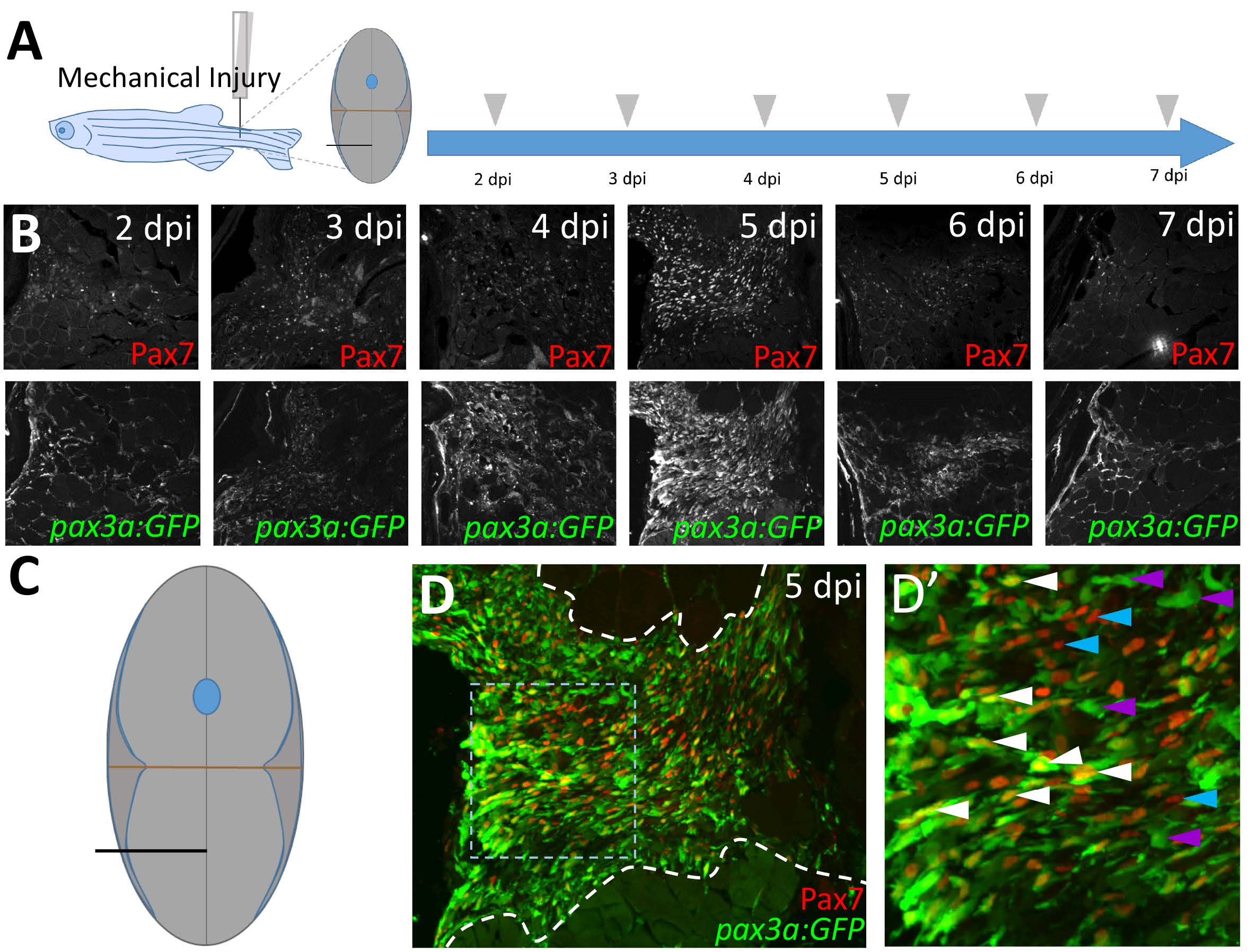
pax3a:GFP expression after injury is similar to that of Pax7, and pax3a:GFP and Pax7 are co-expressed in many cells at the peak of the satellite-like cell response. (A) Schematic of experimental procedure, in which mechanical injury is performed by a single needlestick into tail skeletal muscle. The approximate position of needle-stick injury in the ventral myotome is indicated in cross-sectional view. Tissue was collected at 2, 3, 4, 5, 6, and 7 dpi. (B) The highest number of both pax3a:GFP-positive and Pax7-positive satellite-like cells are detected at 5 dpi in this experiment. Refractive spot in the 7 dpi image is a staining artifact. (C) Schematic representation of ventral needle-stick injury site (black line). (D) Robust Pax7 and pax3a:GFP expression is detected at 5 dpi. Site of tissue repair is located between dashed white lines. (D’) Magnified view of boxed region in D. Satellite-like cells that are both pax3a:GFP and Pax7-positive (white arrowheads), pax3a:GFP-positive (purple arrowheads), and Pax7-positive (blue arrowheads) are indicated.
Reprinted from Developmental Biology, 424(2), Berberoglu, M.A., Gallagher, T.L., Morrow, Z.T., Talbot, J.C., Hromowyk, K.J., Tenente, I.M., Langenau, D.M., Amacher, S.L., Satellite-like cells contribute to pax7-dependent skeletal muscle repair in adult zebrafish, 162-180, Copyright (2017) with permission from Elsevier. Full text @ Dev. Biol.