
Fig. S3
- ID
- ZDB-IMAGE-170509-12
- Publication
- Pradhan et al., 2017 - FGF signaling enforces cardiac chamber identity in the developing ventricle
- All Figures
- Figures for Pradhan et al., 2017
|
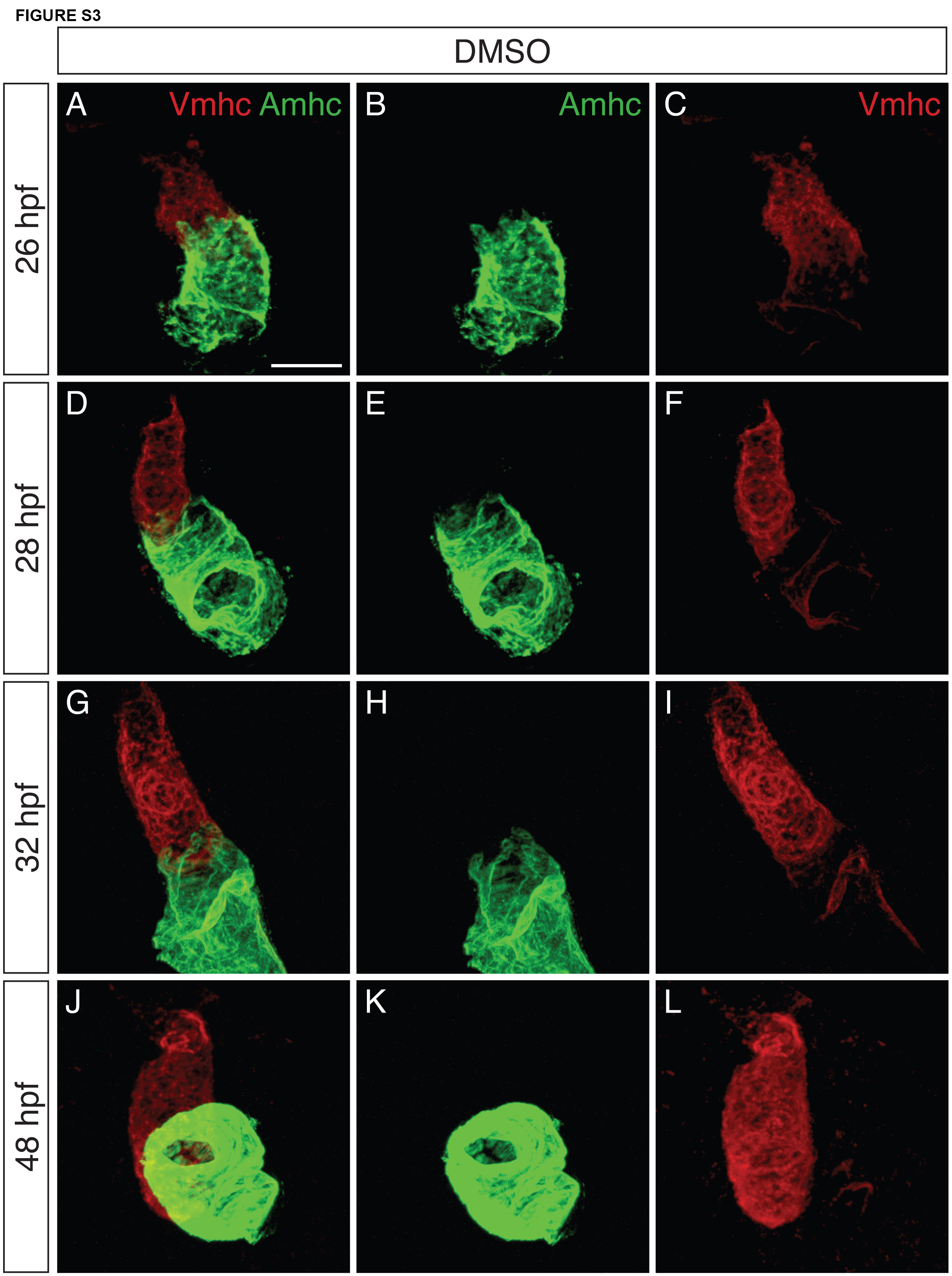
Fig. S3
Vmhc antibody marks expected locations of vmhc expression within the heart. (A-L) Immunofluorescence depicts Vmhc (red) and Amhc (green) localization in wildtype embryos treated with DMSO at 18 hpf. Images are three-dimensional reconstructions of lateral views at 26-48 hpf, showing both channels (A,D,G,J), green only (B,E,H,K), or red only (C,F,I,L). The Vmhc antibody marks the expected locations of vmhc expression within the heart (n=10 for 26.5 hpf; n=8 for 28 hpf; n=8 for 32 hpf; n=8 for 48 hpf). Vmhc immunostaining recapitulates expression of the reporter transgene Tg(vmhc:mCherry-NTR) (Zhang et al., 2013), as well as expression of vmhc itself (Fig. 4I-L). Vmhc is robustly detected in the ventricle, with low levels in the atrium that diminish over time, as has been previously shown for expression of vmhc (Fig. 4I-L) (Yelon et al., 1999). Scale bar: 50 μm.