
Fig. S15
- ID
- ZDB-IMAGE-170508-10
- Publication
- Armstrong et al., 2017 - Shh promotes direct interactions between epidermal cells and osteoblast progenitors to shape regenerated zebrafish bone
- All Figures
- Figures for Armstrong et al., 2017
|
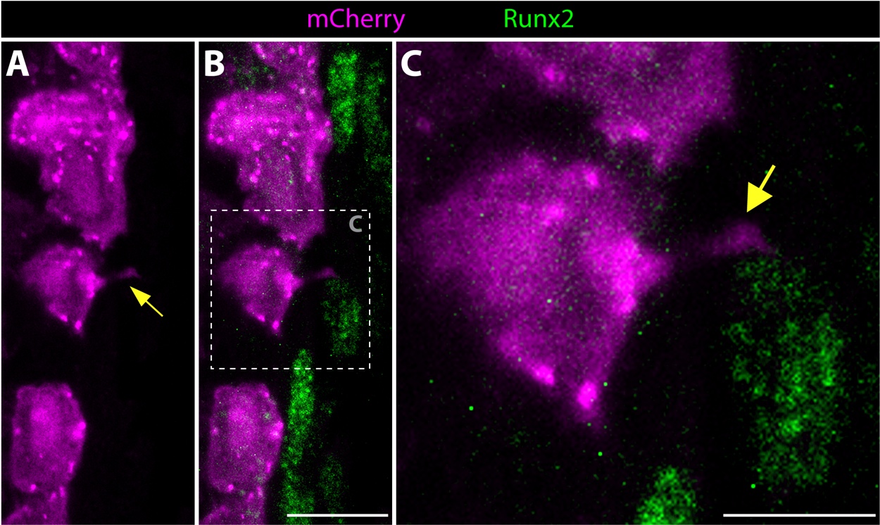
Fig. S15
Distal basal epidermis, fluorescently-labeled using mosaic transgenic zebrafish, extend cellular projections that contact neighboring Runx2+ osteoblast progenitor cells. (A-C) An immunostained distal longitudinal fin section containing mosaic-labeled epidermal cells from a Tg(dusp6:Cre-ERT2, EAB:EGFP-FlEx-mCherry) 96 hpa regenerating fish showing mCherry (magenta) and Runx2 (green) protein. The image is an ~1 μm thick optical section from a confocal stack. Yellow arrows point to a basal epidermal cellular projection that contacts a neighboring pOb. The dashed box in B indicates the zoomed region shown in C. Scale bars are 10 μm (panel B) and 5 μm (panel C).