
Fig. 5
- ID
- ZDB-IMAGE-170425-7
- Publication
- Delfino-Machín et al., 2017 - Sox10 contributes to the balance of fate choice in dorsal root ganglion progenitors
- All Figures
- Figures for Delfino-Machín et al., 2017
|
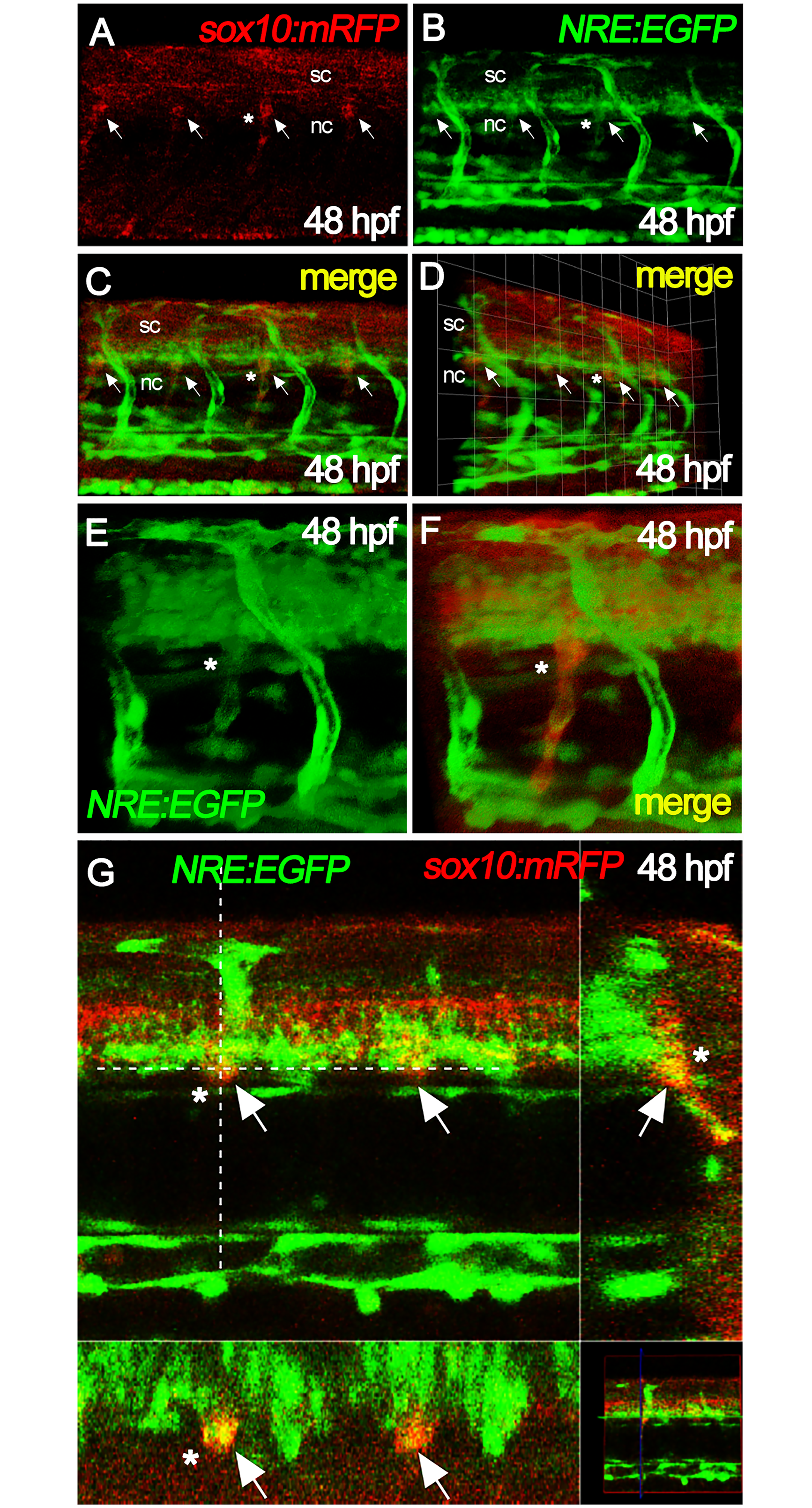
Fig. 5
Notch signaling is active in nascent DRGs.
Neural crest cells in the DRGs of 48 hpf embryos (arrows) were readily identified by expression of mRFP reporter in sox10:mRFP fish (A). These overlapped with eGFP expression from the 12xNRE:eGFP (Notch signaling) reporter (B), as shown in superimposed image (C and D). Close-ups of individual DRGs labelled with asterisk are shown in panels E and F. Panel G shows DRGs (white arrows, yellow signals) as single plane acquisitions, in lateral view (top left), transversal view (top right), dorsal view (bottom left) and mini global view (bottom right). Note that in all panels eGFP is also strongly visible in blood vessels, as expected for the Notch reporter. nc, notochord; sc, spinal cord.