
Image
|
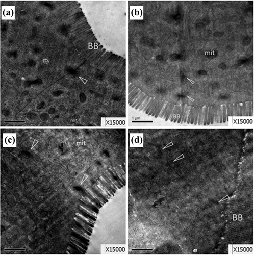
Figure Caption
Fig. 5
Representative TEM images showing small intestine ultrastructure from control females (a) and males (b), and 0.5 μM PFOS-exposed females (c) and males (d). In all females treated with PFOS (c), shrunk or swelling microvilli appeared alternately on the brush border (BB). In all males treated with PFOS, epithelium microvilli disconnected and fell off to intestinal lumen (d). Tight junction (▵) between cells was obscure, cytoplasmic matrix were distributed unevenly; n = 5 fish per sex from each group. TEM: transmission electron microscopic; PFOS: perfluorooctanesulfonic acid.
Acknowledgments
This image is the copyrighted work of the attributed author or publisher, and
ZFIN has permission only to display this image to its users.
Additional permissions should be obtained from the applicable author or publisher of the image.
Full text @ Hum. Exp. Toxicol.