
Fig. S8
- ID
- ZDB-IMAGE-170424-25
- Publication
- Yang et al., 2017 - Miconazole protects blood vessels from matrix metalloproteinase 9-dependent rupture and hemorrhage
- All Figures
- Figures for Yang et al., 2017
|
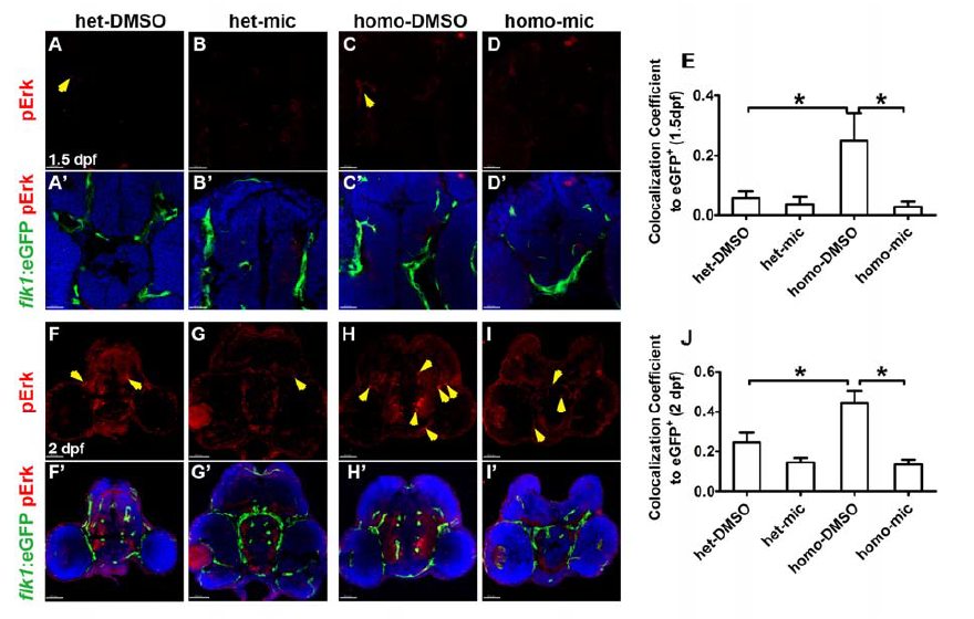
Fig. S8
Miconazole inhibits pErk elevation in the brain vessels of fn40a mutants. Fluorescence immunohistochemistry showed that the phosphorylated Erk1/2 (pErk, red) in Tg(flk1:eGFP) endothelial cells (green) increased in homozygous fn40a mutants (C, C'; H, H') compared with heterozygous siblings in the absence (A, A'; F, F') or presence (B, B'; G, G') of miconazole; and pErk decreased in homozygous mutants after mic treatments (D, D'; I, I'). Tg(flk1:eGFP) (green) was used to label vascular endothelia cells. The colocalization coefficient of pErk and flk1:eGFP to eGFP signals was shown in panel E (1.5 dpf) and panel J (2 dpf), noting that the pErk was up-regulated in mutant endothelium and the elevated pErk was rescued by miconazole treatments. A-D, A'-D', 1.5 dpf, scale bar is 20 μm; F-I, F'-I', 2 dpf, scale bar is 40 μm. DAPI (blue) was used to label nuclei. Yellow arrows indicated the doublepositive signals (pErk/flk1:eGFP). *p <0.05 by one-way ANOVA with Bonferroni's Multiple Comparison Test, n >10 for each zebrafish independent repeat.