
|
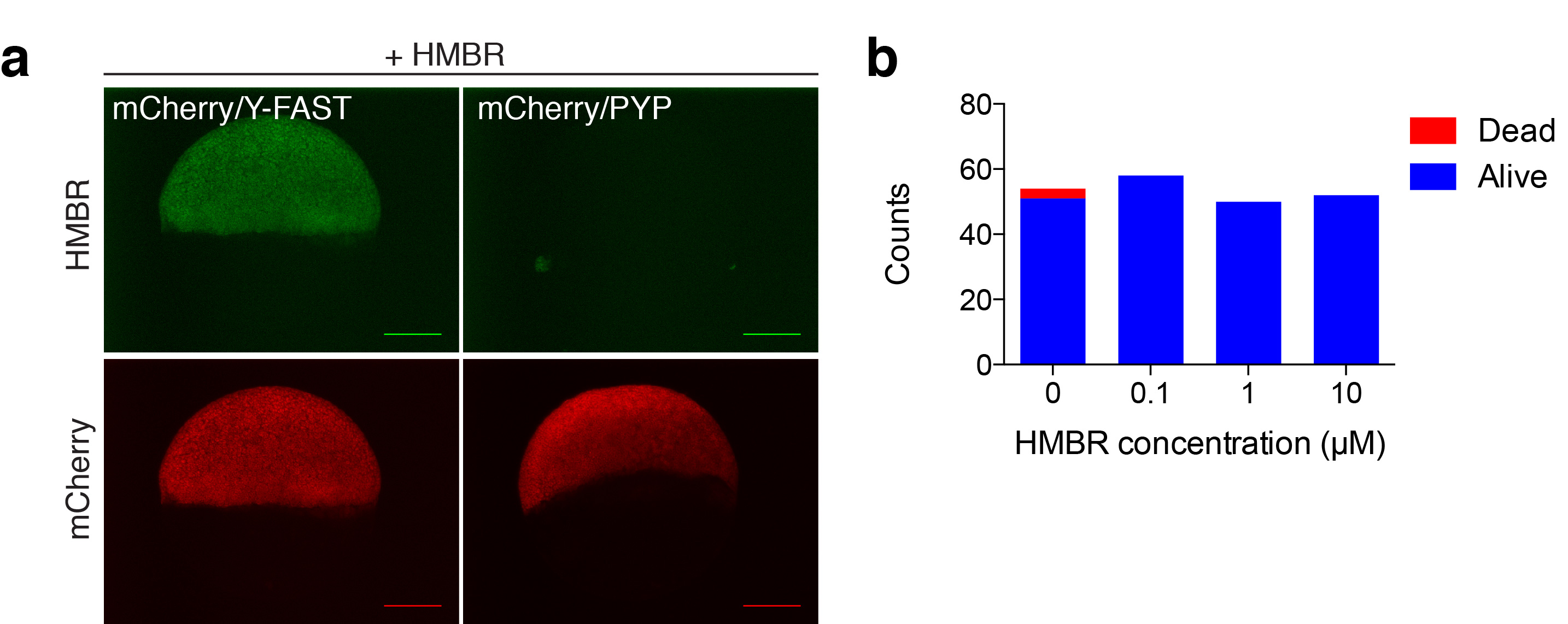
Fig. S16
Labeling of fusion proteins in zebrafish. (a) Spinning-disk confocal micrographs of live zebrafish embryos co-expressing mCherry/Y-FAST or mCherry/PYP labeled with 5 μM HMBR during gastrulation (HMBR channel: Ex/Em 491/525-539 nm, mCherry channel: Ex/Em 561/605-664 nm; scale bars 200 μm). Side-by-side images were recorded using the same settings. (b) Viability of zebrafish embryos incubated with HMBR during development from 50 % epiboly to 24 hpf (19 hours of incubation). The plot shows for various HMBR concentrations the number of embryos that were alive with no morphological defect (blue) or dead (red) at 24 hpf.