
|
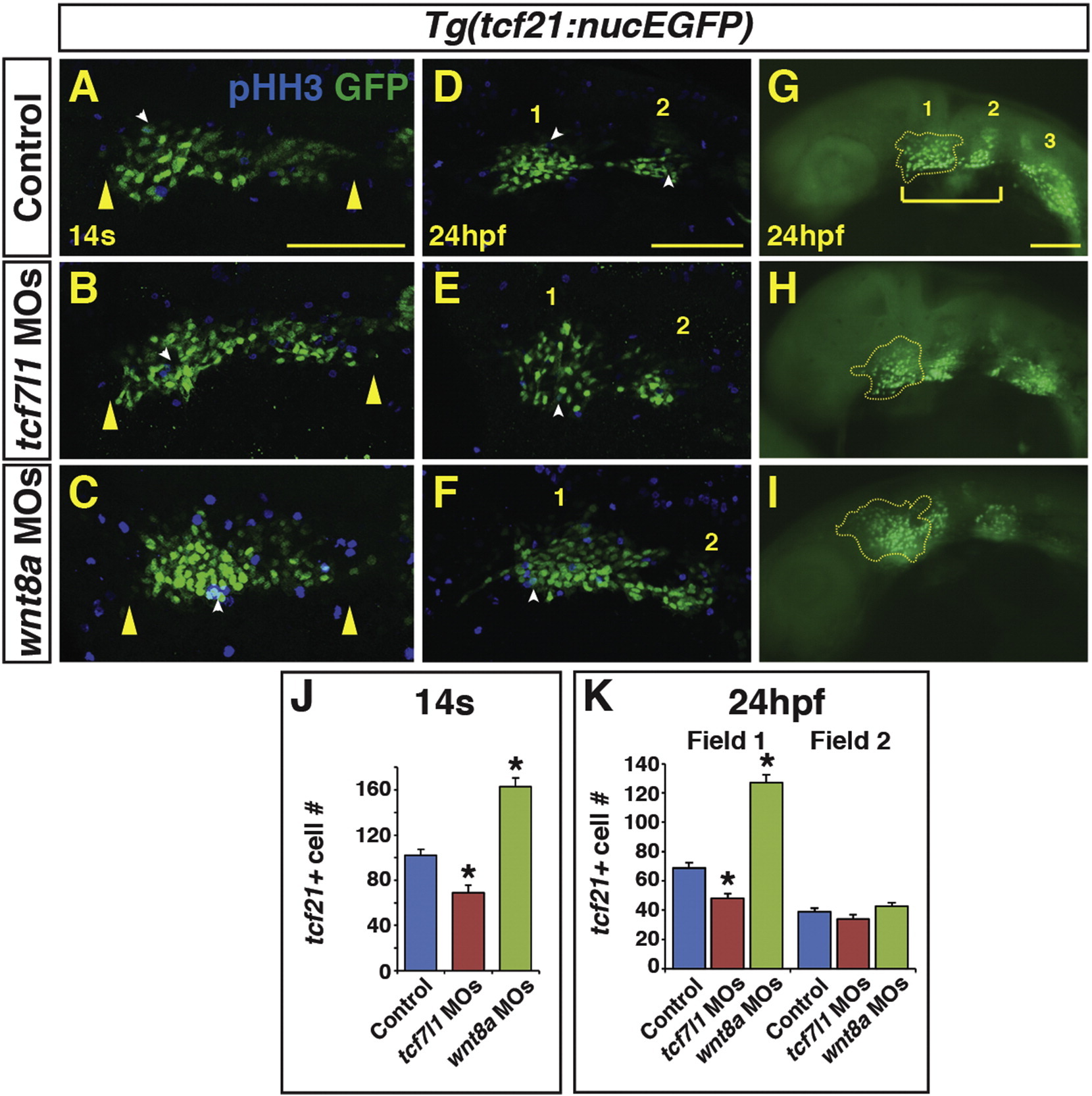
Fig. 5
Endogenous Wnt signaling components restrict tcf21+ PM progenitor development. (A-C) tcf21+ (GFP+) cells within the ALPM of Tg(tcf21:nucEGFP) control, Tcf7l1 depleted, and Wnt8a depleted embryos at the 14 s stage. Images are dorsal views of the left side of the embryos with anterior left. Yellow arrowheads indicate size of field. (D–I) tcf21+ (GFP+) cells within the ALPM of Tg(tcf21:nucEGFP) control, Tcf7l1 depleted, and Wnt8a depleted embryos at 24 hpf. Images are lateral views with anterior left. (E, H) Tcf7l1 depletion produces a loss of the anterior most tcf21+ progenitors (yellow outline). (F, I) Wnt8a depletion results in expansion of the anterior most pharyngeal tcf21+ progenitors. White arrowheads in A–F indicate overlap between GFP+ and phospho-histone H3 (pHH3)+ cells (see Supplementary material – Fig. S3 for analysis of pHH3). Bracket in G indicates the anterior tcf21+ field of cells that give rise to the 1st and 2nd arch muscles. Numbers 1 and 2 in D-G indicate the two anterior tcf21+ fields of cells. Number 3 in G indicates the more posterior tcf21+ population that gives rise to PM in arches 3–7 ( Nagelberg et al., 2015). Images A–F are confocal images. (J) Graph depicting quantification of tcf21+ progenitors at the 14 s stage. Tg(tcf21:nucEGFP) control n = 15, Tcf7l1 depleted n = 10, and Wnt8a depleted embryos n = 10. (K) Graph depicting quantification of tcf21+ progenitors at the 24hpf. Tg(tcf21:nucEGFP) control n = 10, Tcf7l1 depleted n = 13, and Wnt8a depleted embryos n = 10. Cell counts are from confocal images of the tcf21+ field on one side of the embryo. There is a significant decrease in tcf21+ progenitors with Tcf7l1 depletion and conversely a significant increase in tcf21+ progenitors following Wnt8a depletion in the total field at the 14 s stage and anterior field at 24 hpf. Scale bars indicates 100 μm. (For interpretation of the references to color in this figure legend, the reader is referred to the web version of this article.)
Reprinted from Mechanisms of Development, 143, Mandal, A., Holowiecki, A., Song, Y.C., Waxman, J.S., Wnt signaling balances specification of the cardiac and pharyngeal muscle fields, 32-41, Copyright (2017) with permission from Elsevier. Full text @ Mech. Dev.