
Fig. 10
- ID
- ZDB-IMAGE-170413-43
- Publication
- Torres-Paz et al., 2014 - Olfactory sensory system develops from coordinated movements within the neural plate
- All Figures
- Figures for Torres-Paz et al., 2014
|
Fig. 10
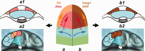
The olfactory sensory system develops as an integrated unit. Center: Schematic of anterior neural plate during early somitogenesis with left side (a) representing the current model with the central olfactory field (1) (telencephalon, red, red arrow) inducing (small red arrows) the peripheral olfactory field (OP, orange, orange arrow) secondarily (2) from ectoderm. Cross-section of forming neural tube at 2 ss (a1) and 20 ss (a2) showing visual field cells (blue) and telencephalic field (red) and the OP (orange). On right (b) the central olfactory field + peripheral olfactory fields (brown) are induced together (1,1) as a common field with shared morphogenetic movements. Cross-section of forming neural tube at 2 ss (b1) and 20 ss (b2) with visual field cells (blue) and a shared central olfactory field + peripheral olfactory fields (brown). Blue shape and arrows represent movements of optic vesicle field during morphogenesis. a2, b2) frontal view of head corresponding to 20 ss. a1, a2, b1, b2 modified from (Whitlock, 2008).