
Image
|
Figure Caption
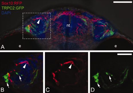
Fig. 9
Sox10:RFP positive neural crest does not contribute to TRPC2:GFP positive microvillar neuronal populations within the olfactory sensory epithelium. A: Whole-mount head at 55 hours postfertilization viewed from ventral. Boxed area indicates region of interest shown in B–D. B: Expression of Sox10:RFP (red, arrowhead) and TRPC2:GFP (green, arrows) in the OP. B: TRPC2:GFP (arrows) in the olfactory organ. C: Expression of Sox10:RFP (arrowhead) in the olfactory organ. B–D: Images are 3 µm optical sections. GFP, green fluorescent protein; RFP, red fluorescent protein. Scale bars = 50 µm in A; 25 µm in B–D.
Acknowledgments
This image is the copyrighted work of the attributed author or publisher, and
ZFIN has permission only to display this image to its users.
Additional permissions should be obtained from the applicable author or publisher of the image.
Full text @ Dev. Dyn.