
|
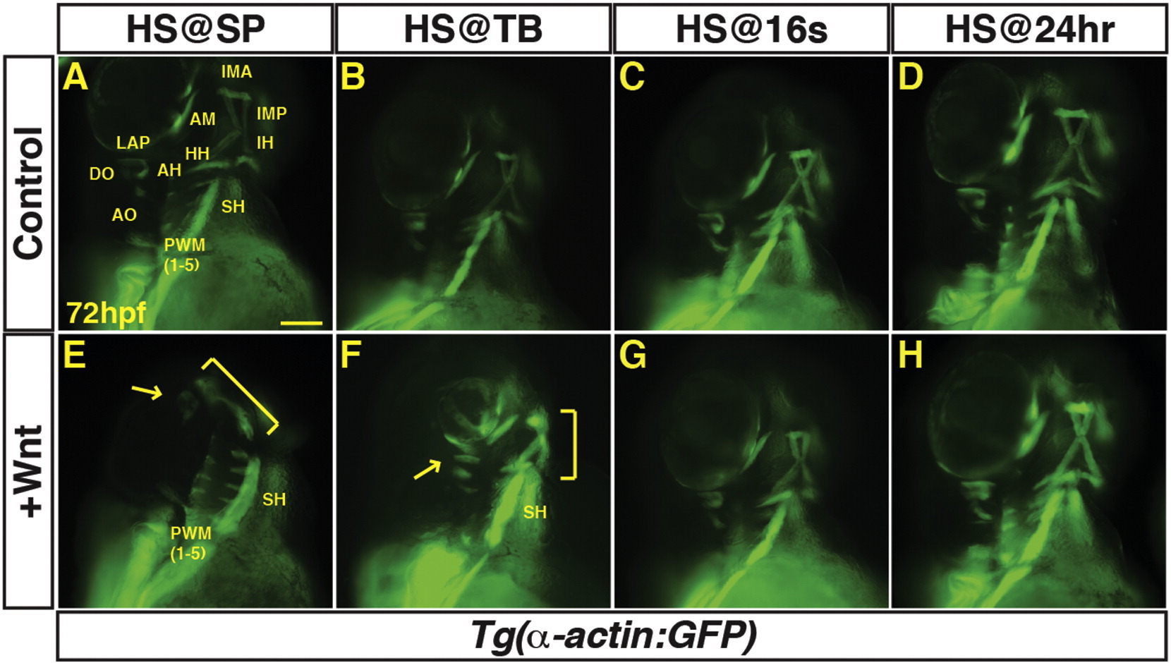
Fig. 1
Excess Wnt signaling inhibits PM development during gastrulation. (A–P) Tg(α-actin:GFP) control sibling embryos and embryos with increased Wnt signaling (hsp70l:wnt8a-GFP+). (A, E) After increasing Wnt signaling from heat-shock (HS) at the SP stage, the EOMs, dorsal 1st and 2nd arch PMs are lost (arrow), while the ventral 1st and 2nd arch PMs are reduced (bracket). PW muscles are also reduced, while the SH was overtly unaffected. (B, F) After increasing Wnt signaling at the TB stage, there is a similar but less severe loss of EOMs, 1st and 2nd arch PMs (dorsal PMs – arrow; ventral PMs bracket). (C, G, D, H) Wnt signaling does not overtly affect PM development at later stages. Muscle nomenclature used is from ( Schilling and Kimmel, 1997). 1st arch muscles - intermandibularis anterior (IMA), intermandibularis posterior (IMP), adductor mandibulae (AM), levator arcus palatine (LAP), dilatator opercula (DO). 2nd (hyoid) arch muscles - hyohyal (HH), interhyal (IH), adductor hyomandibulae (AH), adductor opercula (AO), and levator opercula (LO). Embryos are at 72 hpf. Images are anterior ventro-lateral views. Anterior is up in all images. Heat-shock (HS). > 15 embryos were examined for each condition. Scale bar indicates 100 μm.
Reprinted from Mechanisms of Development, 143, Mandal, A., Holowiecki, A., Song, Y.C., Waxman, J.S., Wnt signaling balances specification of the cardiac and pharyngeal muscle fields, 32-41, Copyright (2017) with permission from Elsevier. Full text @ Mech. Dev.