
Fig. 1
- ID
- ZDB-IMAGE-170413-34
- Publication
- Torres-Paz et al., 2014 - Olfactory sensory system develops from coordinated movements within the neural plate
- All Figures
- Figures for Torres-Paz et al., 2014
|
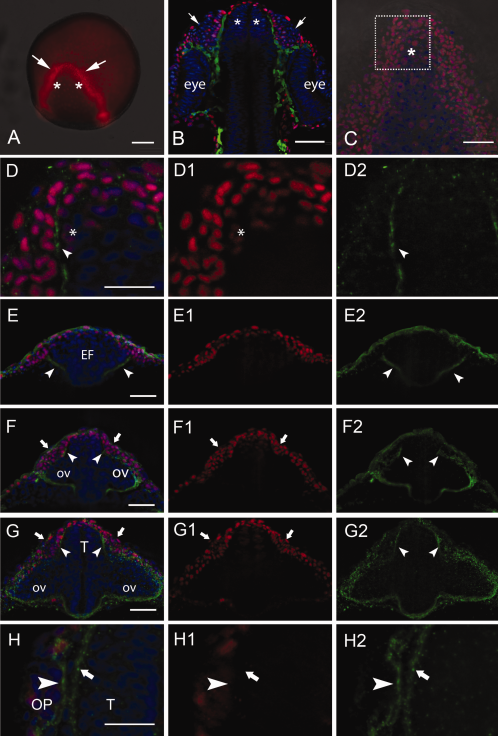
Fig. 1
The Dlx3b protein is expressed in the developing olfactory sensory system. A: The Dlx3b protein (red) is highly expressed in the anterior border of the neural plate at 2 somite stage (ss) in the region giving rise to the olfactory placode (OP) (arrows) and adjacent medial cells (asterisks) will contribute to the telencephalon. B: At 24 hours postfertilization (hpf), the OPs are evident (arrows) adjacent to the telencephalon (asterisks). Cells in green are Sox10:GFP positive neural crest cells separating the OPs from the telencephalon. C,D: Whole-mount embryos, dorsal view, anterior toward top of page. Dlx3b protein 2 ss in the anterior neural plate where region of interest (C, boxed area) shows Dlx3b positive nuclei in the border of the neural plate (D, D1, red) with the initiation of laminin immunoreactivity (D, D2, green, arrow). E: At 2 ss, laminin signal is observed in the ventral border of the eye field (E, E2, green, arrowheads). The dorsal olfactory field (E, E1, red) is not divided by laminin expression at this stage. F: At 8 ss, the laminin signal extends dorsally (F, F2, arrowheads) reaching the telencephalic and OP fields (F, F1, red, arrows). G: At 14 ss, laminin immunoreactivity (G, G2, arrowheads) separates telencephalic and OP fields (G, G1, arrows). H: At 20 ss, laminin immunoreactivity is much stronger (H, H2) and is localized to the region of the basal lamina of the OP (H, H2, arrowhead) and telencephalon (H, H2, arrows). All images are whole-mount embryos. A–D, H are dorsal views; E–G, are frontal views. EF, eye field; GFP, green fluorescent protein; ov, optic vesicle, T, telencephalon. Scale bars = 100 μm in A; 50 μm in B,C; 25 μm in D; 50 µm in E–G; 25 μm in H.