
|
Fig. S5
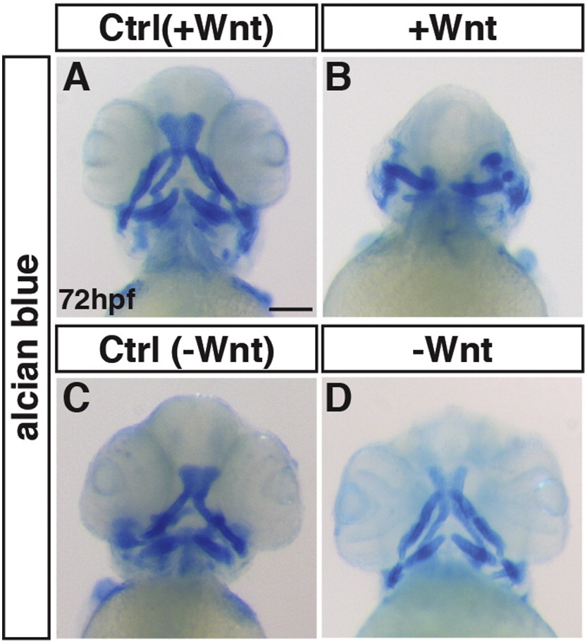
Effects of early Wnt signaling modulation on cranial cartilage. Alcian blue staining at 72 hpf. (A, B) Control sibling embryos and embryos with increased Wnt signaling (hsp70l:wnt8a-GFP+). The anterior neural crest-derived cartilaginous arches are lost and malformed after increased Wnt signaling. (C,D) Control sibling embryos and embryos with decreased Wnt signaling (hsp70l:dkk1-GFP+). The neural crest derived cartilage was minimally affected after loss of Wnt signaling, despite the larger heads. Images are anterior and ventral views. Anterior is up. ≥ 15 embryos were examined per condition. Scale bar indicates 100 μm.
Reprinted from Mechanisms of Development, 143, Mandal, A., Holowiecki, A., Song, Y.C., Waxman, J.S., Wnt signaling balances specification of the cardiac and pharyngeal muscle fields, 32-41, Copyright (2017) with permission from Elsevier. Full text @ Mech. Dev.