
Image
|
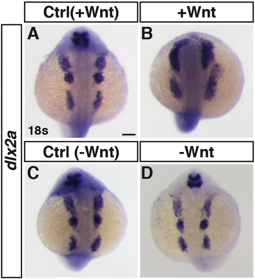
Figure Caption
Fig. S4
Effects of early Wnt signaling modulation on neural crest marker expression. ISH for dlx2a at 18 s stage. (A,B) Control sibling embryos and embryos with increased Wnt signaling (hsp70l:wnt8a-GFP+). The anterior 1st and 2nd neural crest streams are fused and the 3rd is expanded, consistent with posteriorization of the embryo. (C,D) Control sibling embryos and embryos with decreased Wnt signaling (hsp70l:dkk1-GFP+). The neural crest cells were minimally affected after loss of Wnt signaling. Images are dorsal views. Anterior is up. ≥ 15 embryos were examined per condition. Scale bar indicates 100 μm.
Acknowledgments
This image is the copyrighted work of the attributed author or publisher, and
ZFIN has permission only to display this image to its users.
Additional permissions should be obtained from the applicable author or publisher of the image.
Reprinted from Mechanisms of Development, 143, Mandal, A., Holowiecki, A., Song, Y.C., Waxman, J.S., Wnt signaling balances specification of the cardiac and pharyngeal muscle fields, 32-41, Copyright (2017) with permission from Elsevier. Full text @ Mech. Dev.