
Image
|
Figure Caption
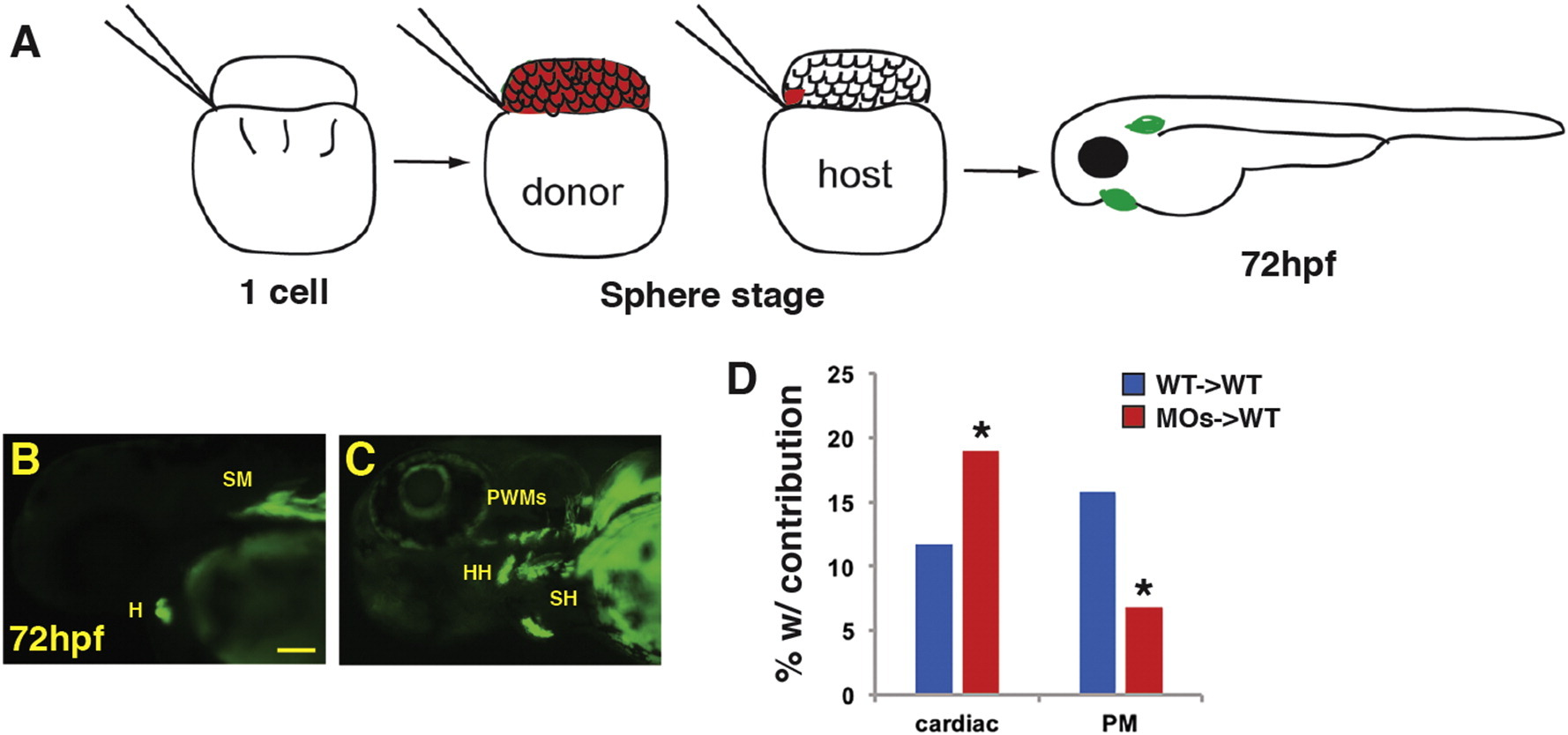
Fig. 9
Wnt signaling cell-autonomously inhibits PM specification. (A) Schematic of blastula cell transplantation method. (B, C) Representative donor cells that incorporated into the heart and PMs at 72 hpf. (D) Graph indicating the frequency of donor cell contribution in embryos to the heart and the PMs. WT donor cells → WT host transplants n = 240. Tcf7l depleted donor cells → WT host embryos n = 221. Tcf7l1 depleted donor cells contributed significantly more to the cardiomyocytes and less frequently to the PMs compared to WT donors. Images are lateral views with anterior leftwards. Scale bar indicates 100 μm.
Acknowledgments
This image is the copyrighted work of the attributed author or publisher, and
ZFIN has permission only to display this image to its users.
Additional permissions should be obtained from the applicable author or publisher of the image.
Reprinted from Mechanisms of Development, 143, Mandal, A., Holowiecki, A., Song, Y.C., Waxman, J.S., Wnt signaling balances specification of the cardiac and pharyngeal muscle fields, 32-41, Copyright (2017) with permission from Elsevier. Full text @ Mech. Dev.