
IMAGE
Fig. S2
- ID
- ZDB-IMAGE-170321-29
- Publication
- Liu et al., 2017 - Stat3/Cdc25a-dependent cell proliferation promotes embryonic axis extension during zebrafish gastrulation
- All Figures
- Figures for Liu et al., 2017
Image
|
Figure Caption
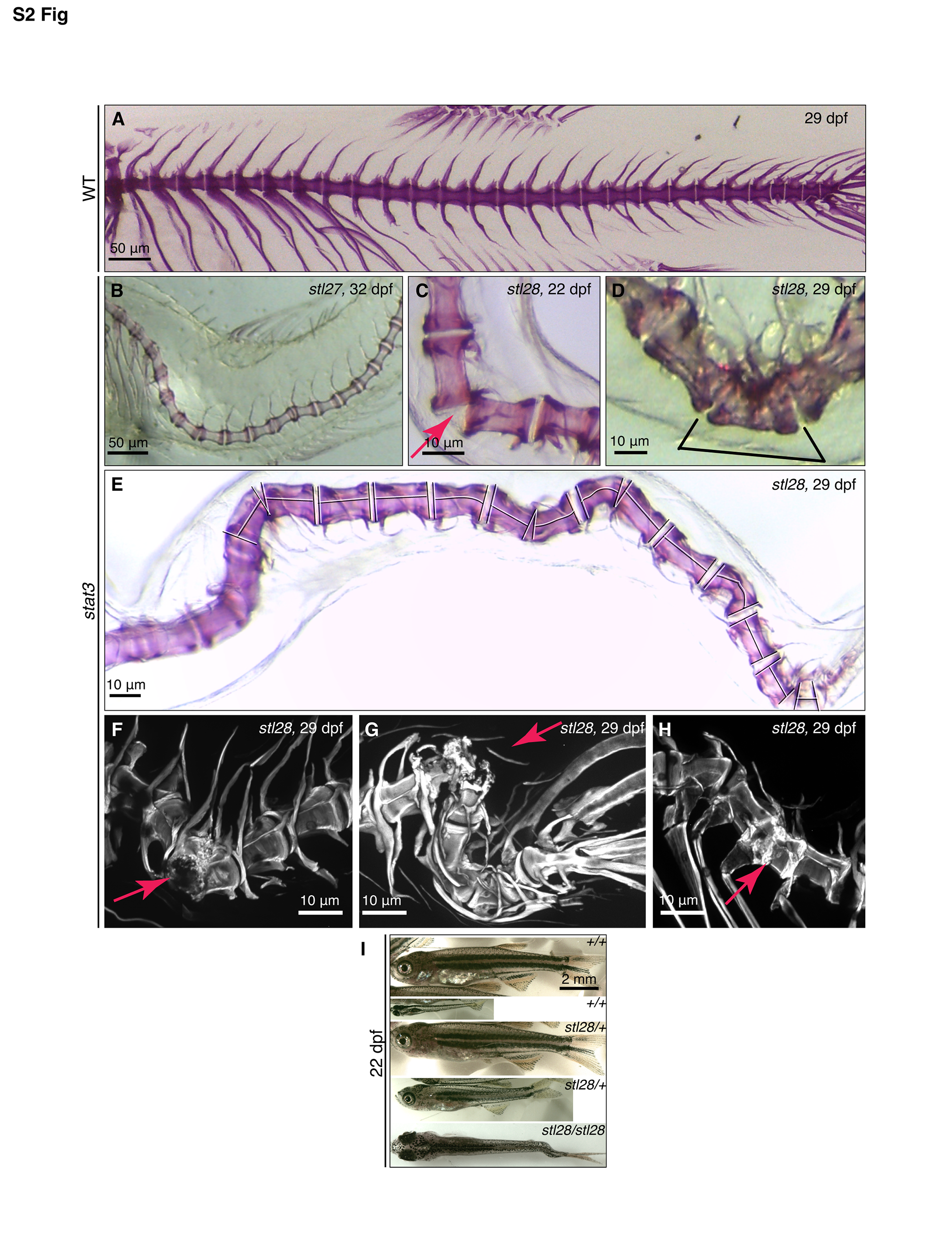
Fig. S2
Various vertebral abnormalities contribute to the scoliosis phenotype in stat3 animals.
(A) Vertebrae of a WT larva at 29 dpf; anterior to the left. (B-H) Images of Alizarin stained skeletons showing various vertebral abnormalities. B and C, normal vertebral body and end plates, tilted intervertebral discs; D and E, bent vertebral body and non-perpendicular end plates; F-H, fractures and extra bony matrix. (I) Variations in larvae body length of stat3 mutant and siblings at 22 dpf.
Acknowledgments
This image is the copyrighted work of the attributed author or publisher, and
ZFIN has permission only to display this image to its users.
Additional permissions should be obtained from the applicable author or publisher of the image.
Full text @ PLoS Genet.