
Fig. 5
- ID
- ZDB-IMAGE-170321-24
- Antibodies
- Publication
- Liu et al., 2017 - Stat3/Cdc25a-dependent cell proliferation promotes embryonic axis extension during zebrafish gastrulation
- All Figures
- Figures for Liu et al., 2017
|
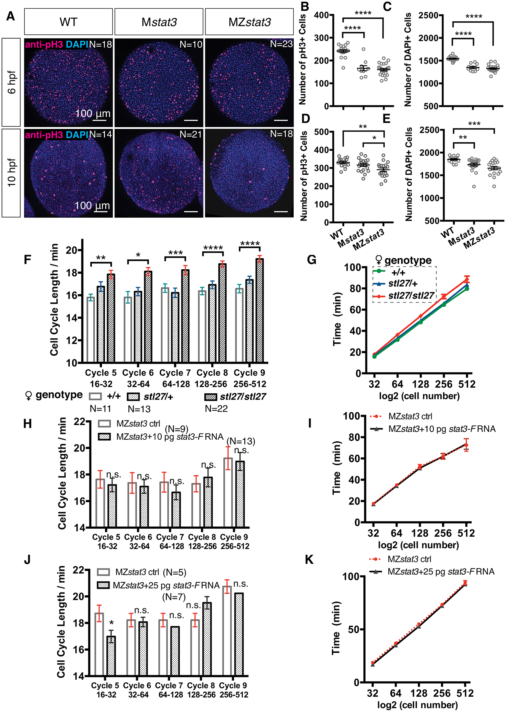
Fig. 5
Stat3 promotes cell cycle progression during zebrafish embryogenesis.
(A) Immunofluorescent anti-pH3 labeling of proliferating cells (red) and DAPI labeling all nuclei (blue) in WT, Mstat3, and MZstat3 embryos at 6 hpf (animal view) and 10 hpf (dorsal view). (B, D) Quantification of mitotic cell number at 6 and 10 hpf. (C, E) Quantification of total cell number at 6 and 10 hpf. (F and G) Average length of each cell cycle (F) and timing of mitosis (G) from Cycle 5 to Cycle 9 in embryos from WT, stat3stl27/+, and stat3stl27/+ females. (H-K) Analyses of cell divisions from Cycle 5 to Cycle 9 in 10 pg (H and I) and 25 pg (J and K) stat3-F injected MZstat3 embryos. *p<0.05, **p<0.01, ***p<0.001, ****p<0.0001, n.s. = non-significant, error bars = SEM. See also S5 Fig.