
|
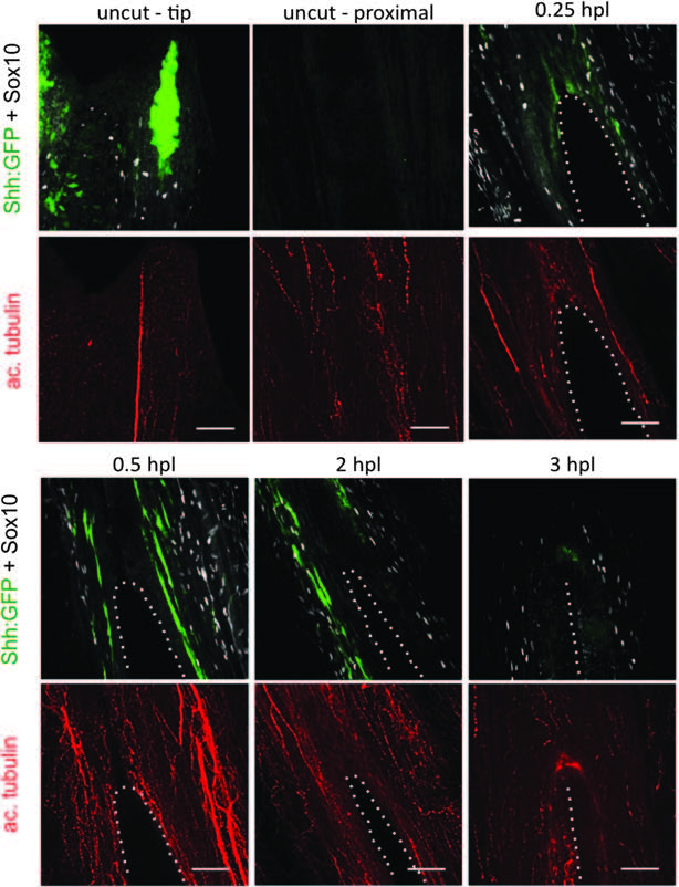
Fig. S1
Sox10 and Shh are activated shortly after lesion. Immunofluorescence staining for immature SC marker, Sox10 (white), Shh-expressing cells (anti-GFP, Shh:GFP fish) (green), and the axonal marker, acetylated tubulin (red). A lesion was performed in between the rays. As soon as 0.25 hpl, Sox10-positive and Shhexpressing cells are detected surrounding the lesion. Once the wound is healed (3 hpl), Shh-expressing cells are no longer detectable, whereas several Sox10-positive cells continue to set along the wound. Scale bars = 50 µM. Dotted lines represent the hole generated by the lesion. GFP, green fluorescent protein; hpl, hours postlesion; SC, Schwann cell.