
|
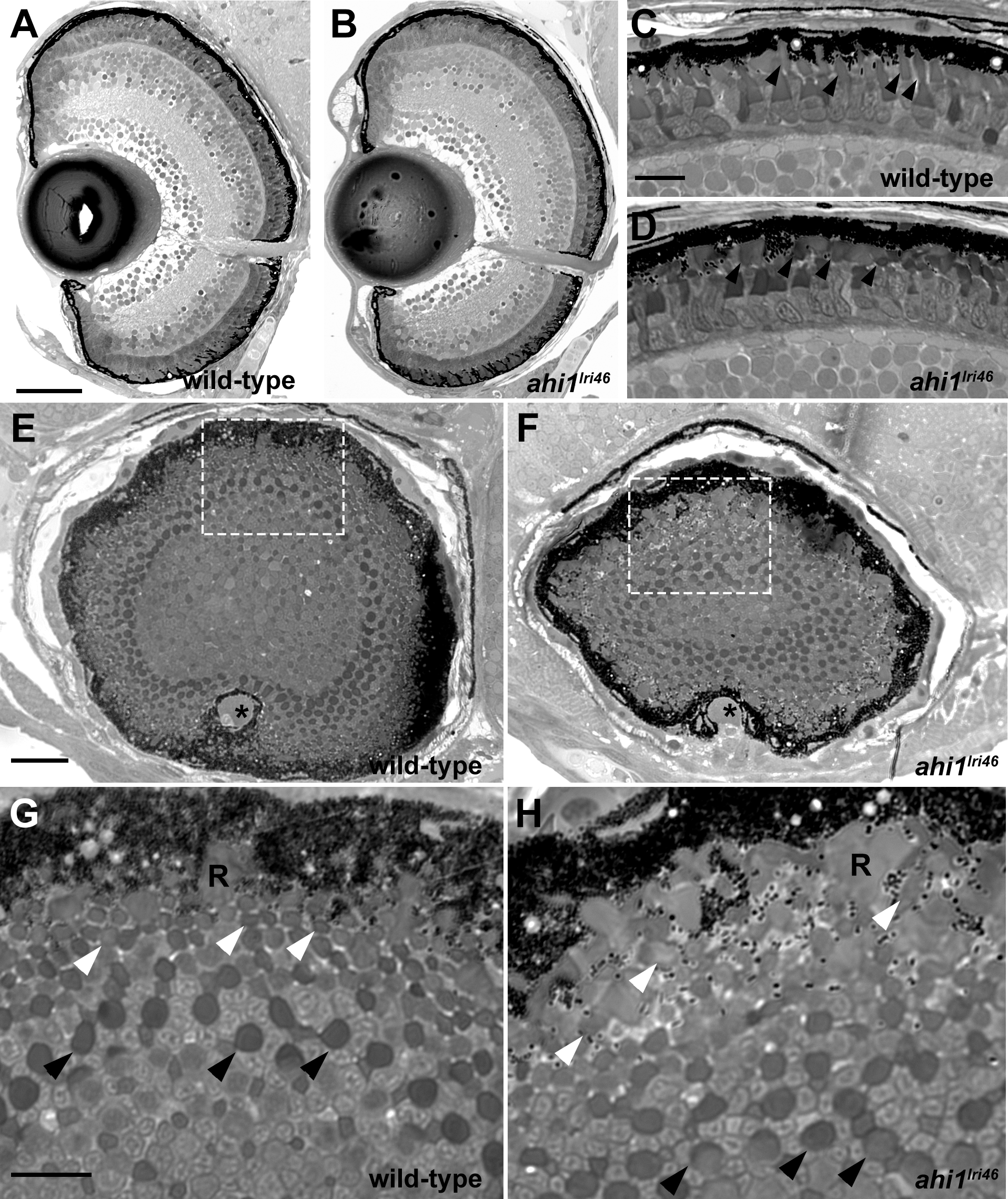
Fig. 3
Histologic survey of retinas of 5 dpf wild-type and ahi1lri46 larvae (A–D) Transverse sections of 5 dpf wild-type and ahi1lri46 eyes. Photoreceptor outer segments (arrowheads) were shorter and less organized in ahi1lri46 mutants. (E–H) Sagittal sections of wild-type and ahi1lri46 eyes, taken at the posterior pole just dorsal to the optic nerve (*). Indicated regions (white dashed boxes) in (E, F) are shown at higher magnification in (G, H). Black arrows indicate the larger caliber, more proximal UV cone outer segments. White arrows indicate the smaller caliber, more distal red, green, and blue cone outer segments. R, rod outer segments. Scale bars: 50 μm (A, B), 10 μm (C, D, G, H) and 25 μm (E, F).