
|
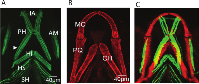
Fig. 1
Representative images of the musculoskeletal elements of the zebrafish lower jaw at 5 dpf. Representative confocal stacks of the lower jaw of 5dpf larvae all shown with anterior to top (A) Immunostaining for A4.1025 which stains all skeletal myosin (B) Immunostaining for Type II collagen which marks all cartilage (C) Stack from a live larva expressing the transgenic reporters col2a1:mCherry marking cartilage (red) and smyhc:GFP slow muscle (green). IA: intermandibularis anterior, PH: protractor hyoideus, AM:adductor mandibulae, HI: hyoideus inferior, HI: hyoideus superior, CH: sternohyoideus, MC: Meckel's cartilage, PQ: Palatoquadrate, CH: ceratohyal.