
IMAGE
Fig. 1 S1
- ID
- ZDB-IMAGE-170222-58
- Publication
- Tenente et al., 2017 - Myogenic regulatory transcription factors regulate growth in rhabdomyosarcoma
- All Figures
- Figures for Tenente et al., 2017
Image
|
Figure Caption
Fig. 1 S1
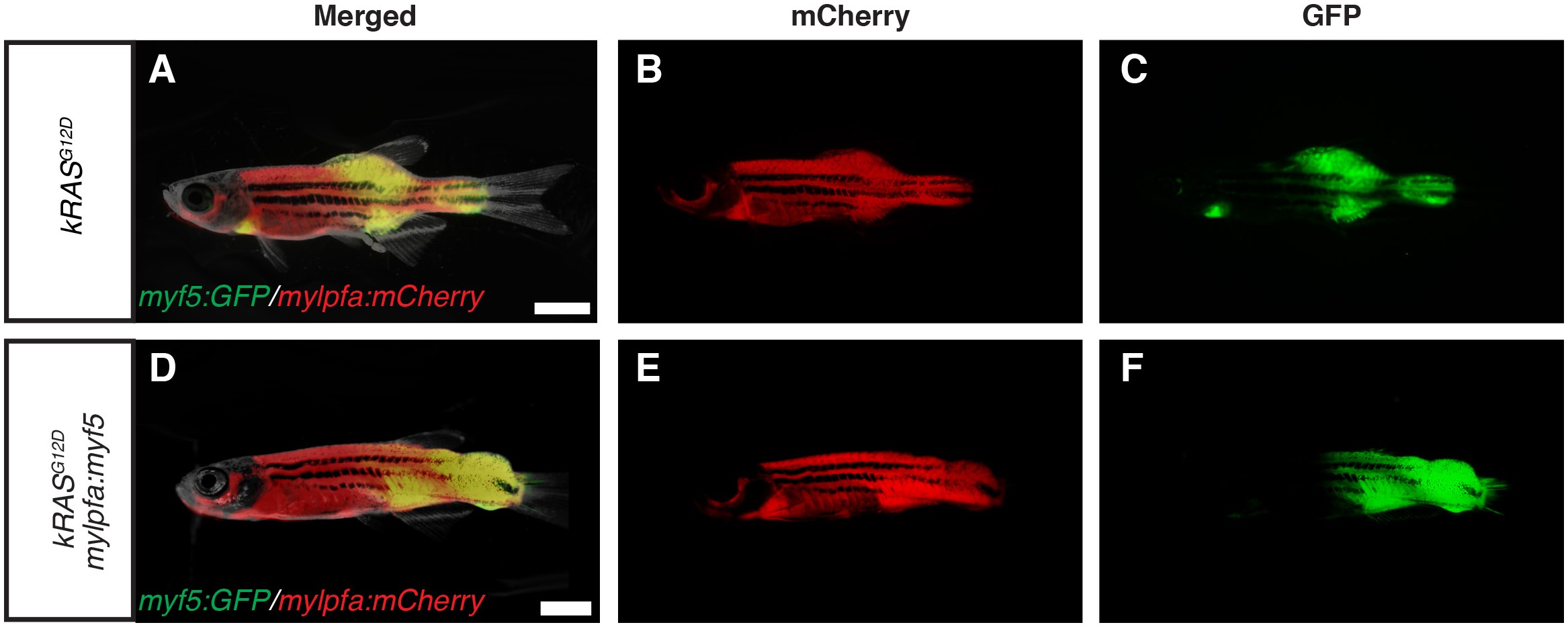
Fluorescence images of primary ERMS developing in stable transgenic myf5:GFP/mylpfa:mCherry zebrafish.
Images of the same representative rag2:kRASG12D –alone (A–C) and rag2:kRASG12D; mylpfa:myf5 (D–F) zebrafish shown in Figure 1A and D, respectively. (A,D) merged (brightfield, GFP and mCherry) image. (B,E) mCherry image. (C,F) GFP image. Scale bars equal 2 mm.
Acknowledgments
This image is the copyrighted work of the attributed author or publisher, and
ZFIN has permission only to display this image to its users.
Additional permissions should be obtained from the applicable author or publisher of the image.
Full text @ Elife