
Fig. 7
- ID
- ZDB-IMAGE-170222-36
- Publication
- Fontenas et al., 2016 - Neuronal Ndrg4 Is Essential for Nodes of Ranvier Organization in Zebrafish
- All Figures
- Figures for Fontenas et al., 2016
|
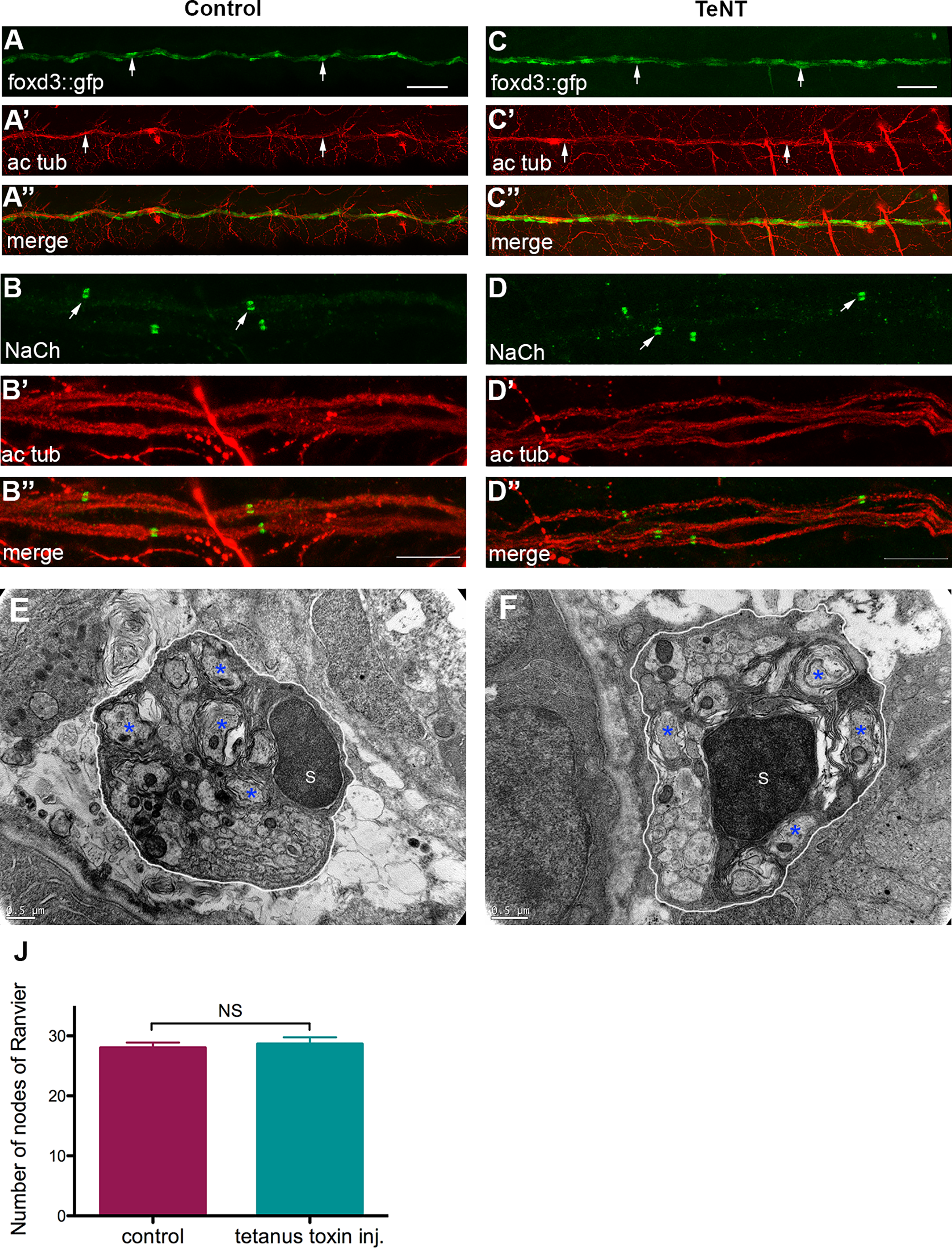
Fig. 7
Tetanus toxin injection does not impair sodium channel clustering and myelination in the PNS.
(A) Lateral view of a control foxd3::GFP embryo at 3 dpf. Arrows indicate SCs along the PLLn. (A') Acetylated tubulin expression in the same control embryo at 3 dpf. Arrows show the PLLn axons. (A'') Merge of (A) and (A’). (C) Lateral view of a TeNT injected foxd3::GFP embryo at 3 dpf. Arrows indicate SCs along the PLLn. (C') Acetylated tubulin expression in the same TeNT injected embryo at 3 dpf. Arrows indicate the PLLn axons. (C'') Merge of (C) and (C'). Scale bars = 50μm. Sodium channel labeling in control (B) and TeNT injected embryo (D) and their corresponding axons of the PLLn, (B') and (D') respectively. (B'') merge of (B) and (B'), (D'') merge of (D) and (D’). Scale bars = 10μm. (E,F) Transmission electron micrographs showing cross-section through (E) control and (F) TeNT injected embryos’ PLLn at 4 dpf. (E) Control PLLn shows an average of 6 myelinated axons (blue asterisks). (F) An average of 6 myelinated axons (blue asterisks) is also observed in the TeNT injected embryo’s PLLn. (S: Schwann cell). Scale bars = 0.5μm. (J) Quantification of the number of nodes seen in the PLLn shows no significant difference between controls and TeNT injected embryos.