
Fig. 4
- ID
- ZDB-IMAGE-170222-33
- Publication
- Fontenas et al., 2016 - Neuronal Ndrg4 Is Essential for Nodes of Ranvier Organization in Zebrafish
- All Figures
- Figures for Fontenas et al., 2016
|
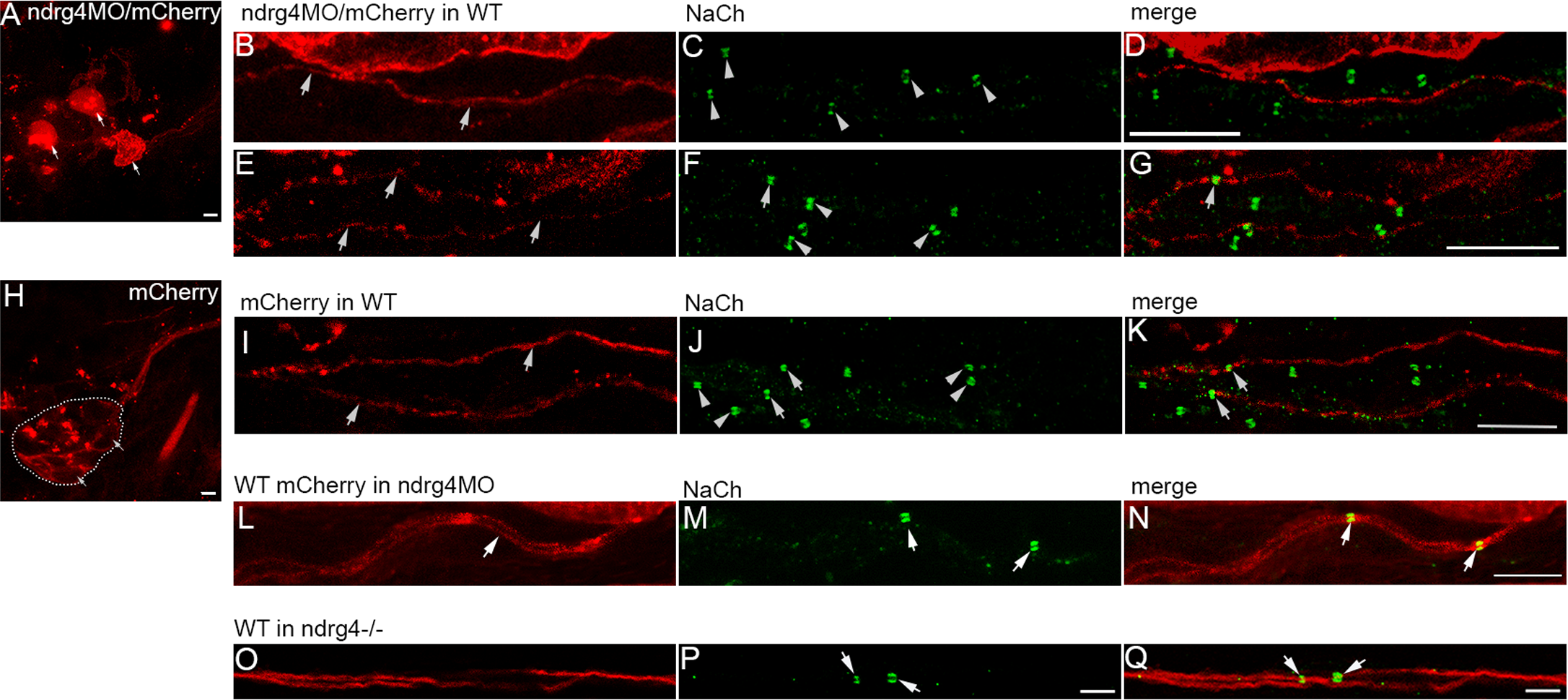
Fig. 4
Chimeric embryos show evidence of ndrg4 requirement in neurons for sodium channel clustering.
(A) ndrg4MO mCherry labeled PLL neurons shown by arrows. (b,e) ndrg4MO mCherry labeled axons of the PLLn in two different chimeric embryos; (c,f) sodium channels along the PLLn of ndrg4MO mCherry labeled (arrow) and of control (arrowheads) axons; (d,g) merge of the two labelings. Sodium channel clustering is absent in ndrg4MO axons (mCherry labeled) while control ones in the same PLLn show normal clustering. (H) Control mCherry labeled neurons indicated by arrows. The dashed line indicates the margin of the PLLg. (I) Control mCherry labeled axons (arrows). (J) Sodium channel clusters along the PLLn in control labeled (arrows) and other non-labeled (arrowheads) axons. (K) Merge of the two labelings. For (a, H) scale bars = 5μm. For (b-G; I-K) scale bars = 10μm. (L-N) WT mCherry labeled axons in ndrg4 morphant embryos are shown in (L, arrow) and the corresponding sodium channels in (M, arrows). Note the clustering of the nodes in the WT labeled axons while the other ndrg4 deficient axons show no sign of sodium channel clustering. (N) Merge of the two labelings. Scale bar = 7 μm. (O-Q) WT mcherry labeled axons in ndrg4-/- are shown in (O) and the corresponding sodium channels in (P, arrows). (Q) Merge of the two labelings showing clustered sodium channels along the WT axons (arrows). Scale bar = 5 μm.