
|
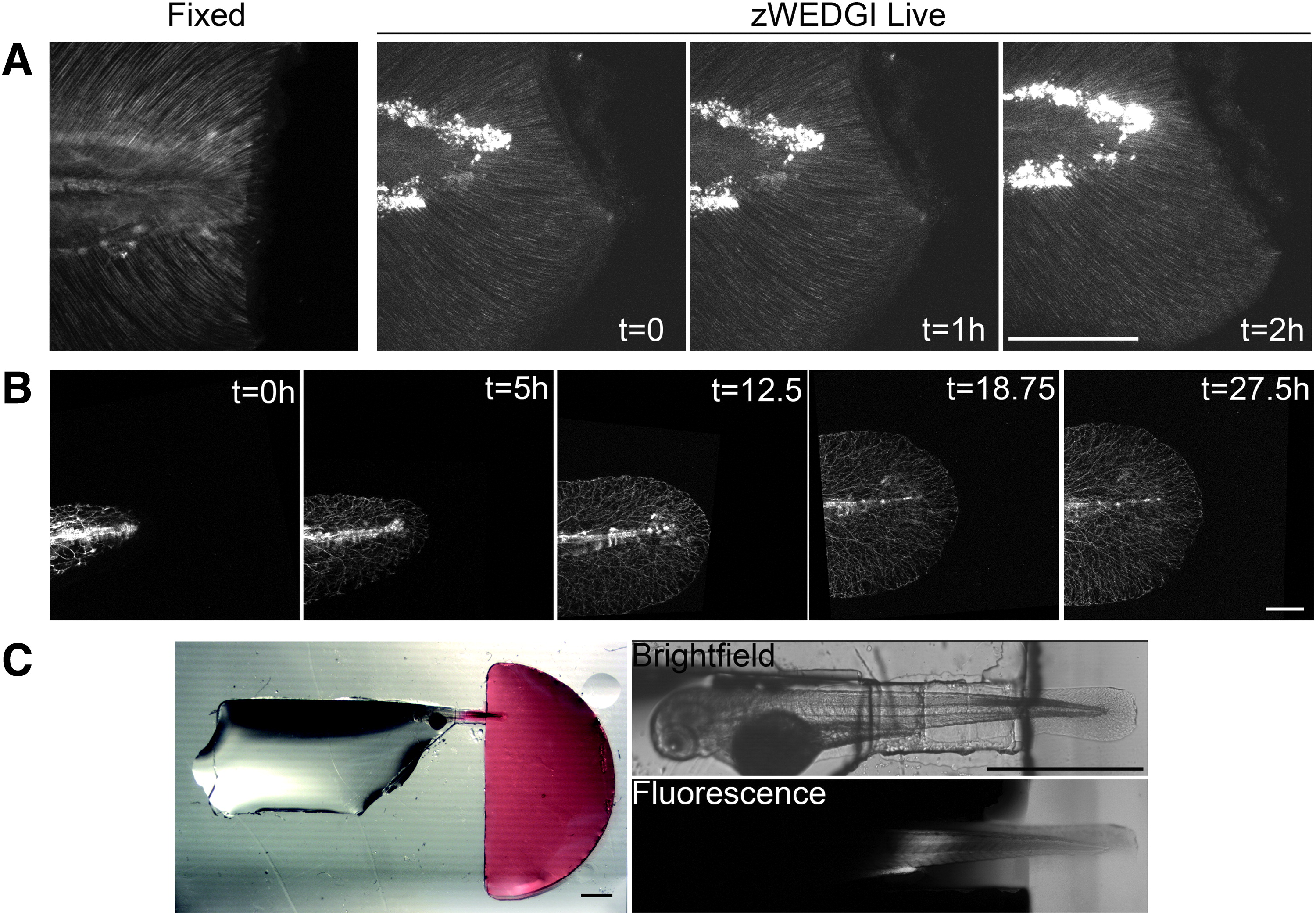
Fig. 4
High-resolution light microscopy of larval tails in zWEDGI. (A) Z-projections of SHG images collected with similar imaging parameters of a fixed sample (single time point) and a live sample mounted in a zWEDGI (multiple time points), demonstrating short time frame imaging immediately after wounding. (B) Confocal long-term time lapse of tail development showing gfp-tagged neuron growth. (C) Dissecting microscope image with accompanying multiphoton image showing isolation of rhodamine 6G (red) to the wounding chamber, 2 h after initial application of the dye to the wounding chamber, illustrating only minor infiltration of dye into the restraining tunnel and levels below detection in the head region. (A, B) Scale bar = 100 μm. (C) Scale bar = 1 mm. SHG, second harmonic generation.