
|
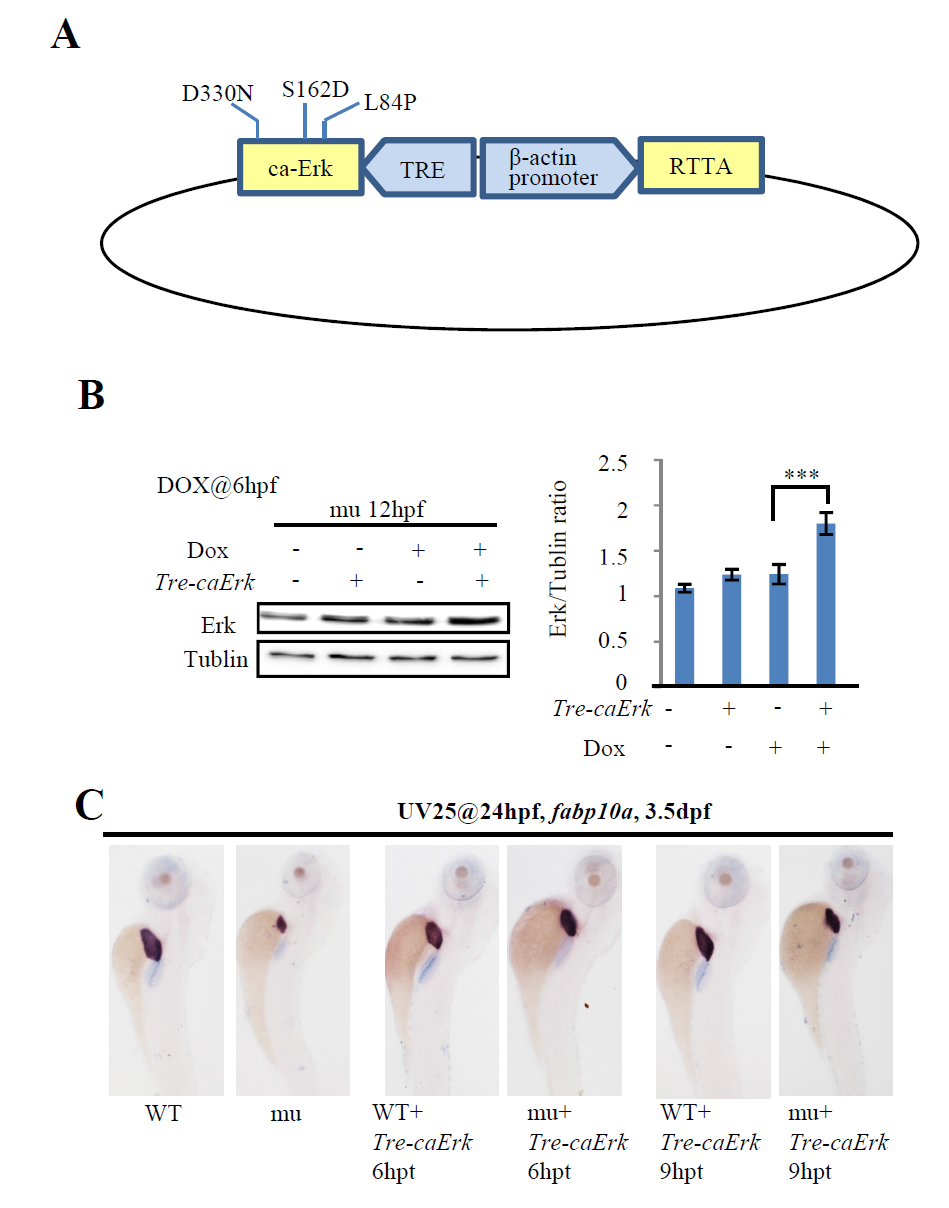
Fig. S6
Construction of the Tre-caErk plasmid.
(A) Schematic drawing showing the structure of the TRE-caErk plasmid. RTTA expression was driven by the β-actin gene promoter. Dox binds to RTTA and the Dox-RTTA complex binds to the TRE promoter to drive the expression of caErk. (B) 10 pgTre-caErk plasmid DNA was injected into one-cell stage maternal-zygotic leg1azju1 embryos (mu). These embryos were treated with Dox at 6 hpf and total protein was harvested at 12 hpf. The protein samples were subjected to western blot analysis. The total Erk versus Tubulin ratios were shown on the right. Dox, doxycycline. Error bar stands for the standard error. ***, p<0.001. Western blot was repeated three times. (C) Images of representative 3.5-dpf embryos after WISH using the fabp10a probe. Embryos was first injected with Tre-caErk plasmid at one-cell stage, then treated with UV25 at 24 hpf and followed by Dox treatment for 6 hours (6 hpt) or 9 hours (9 hpt). After Dox treatment, embryos were transferred to the normal egg water to grow to 3.5 dpf for WISH (n = 20).