
IMAGE
Fig. S8
- ID
- ZDB-IMAGE-170208-23
- Publication
- Bielczyk-Maczyńska et al., 2015 - The Ribosome Biogenesis Protein Nol9 Is Essential for Definitive Hematopoiesis and Pancreas Morphogenesis in Zebrafish
- All Figures
- Figures for Bielczyk-Maczyńska et al., 2015
Image
|
Figure Caption
Fig. S8
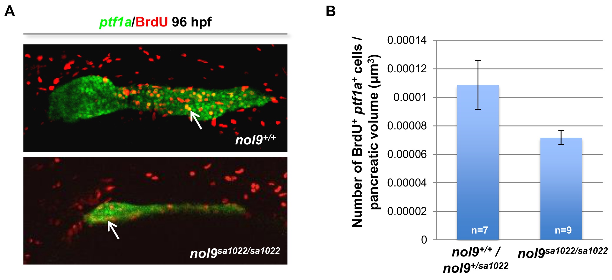
Proliferation of exocrine pancreas cells in nol9sa1022/sa1022 mutants.
(A) Representative confocal images of Tg(ptf1a:EGFP) larvae subjected to BrdU incorporation assay at 96 hpf. Double positive ptf1a+ BrdU+ cells are indicated with arrows. Images are oriented with anterior to the right and dorsal to the top. (B) Average number of ptf1a+ cells which incorporated BrdU, normalized to the volume of ptf1a+ exocrine pancreas, in nol9sa1022/sa1022 (n = 9) and wt (n = 7) larvae at 96 hpf. Data are represented as the mean +/- SEM. Student’s t-test, p = 0.051.
Acknowledgments
This image is the copyrighted work of the attributed author or publisher, and
ZFIN has permission only to display this image to its users.
Additional permissions should be obtained from the applicable author or publisher of the image.
Full text @ PLoS Genet.