
Fig. S7
- ID
- ZDB-IMAGE-170208-22
- Publication
- Bielczyk-Maczyńska et al., 2015 - The Ribosome Biogenesis Protein Nol9 Is Essential for Definitive Hematopoiesis and Pancreas Morphogenesis in Zebrafish
- All Figures
- Figures for Bielczyk-Maczyńska et al., 2015
|
Fig. S7
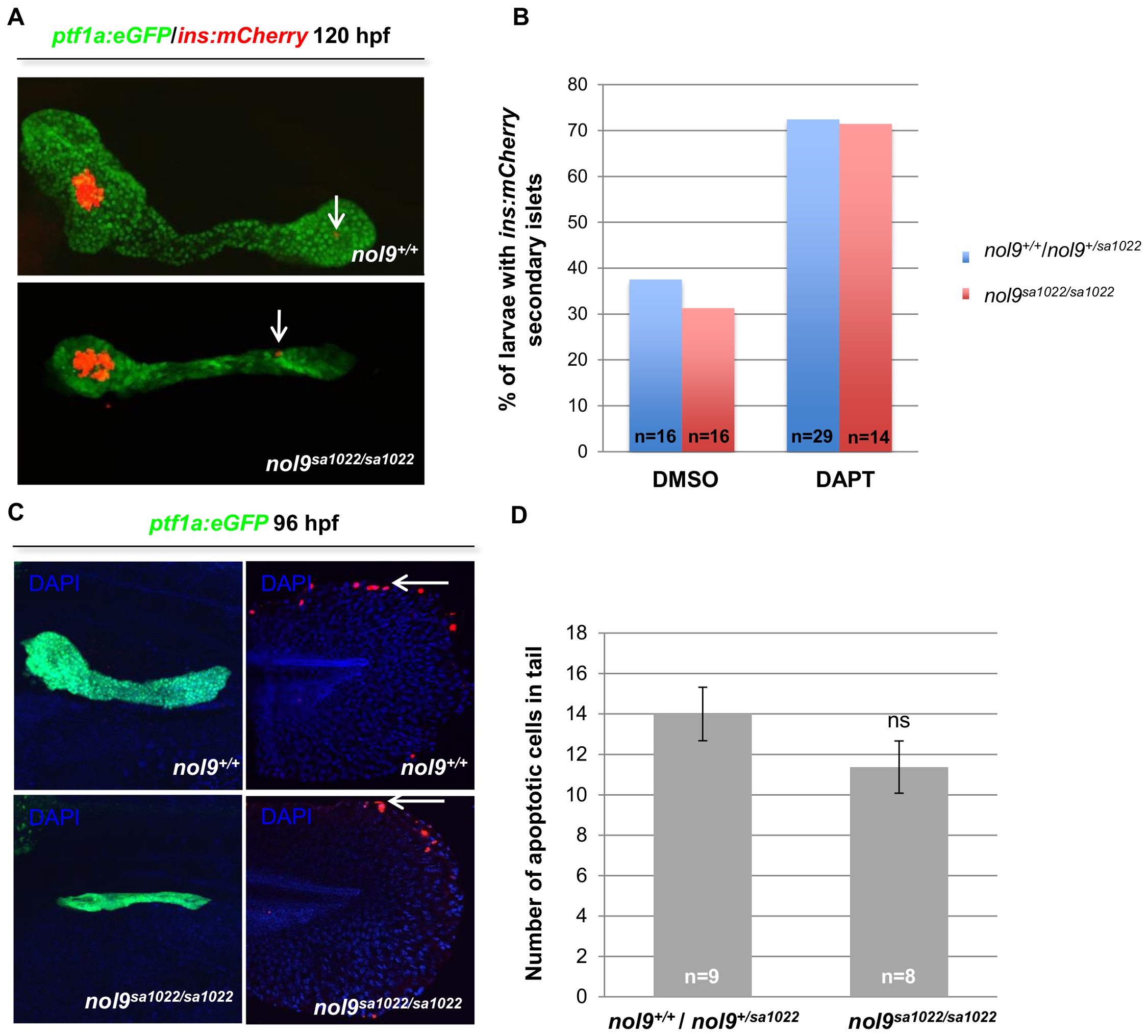
Formation of secondary islets and apoptosis levels in pancreas are unaffected in nol9sa1022/sa1022 mutants.
(A) Representative confocal images of the pancreas of 120 hpf Tg(ptf1a:EGFP;ins:mCherry) larvae showing the presence of ins-expressing secondary islets (arrow) in nol9sa1022/sa1022 mutants and wt siblings. (B) The percentage of Tg(ptf1a:EGFP;ins:mCherry) larvae whose pancreas contained secondary islets, depending on their genotype and previous treatment with either DAPT inhibitor or DMSO (vehicle control) at 120 hpf. The total number of larvae in each group is indicated. (C) Representative confocal images of Tg(ptf1a:EGFP) larvae subjected to TUNEL assay at 96 hpf and co-stained with DAPI. No TMR-labelled apoptotic cells were observed in the ptf1a-expressing exocrine pancreas of nol9sa1022/sa1022 mutants (n = 8) or their wt siblings (n = 9). However, the tails of nol9sa1022/sa1022 mutants and wt siblings contained similar numbers of apoptotic cells (arrow). (D) The mean number of apoptotic cells in the tails of nol9sa1022/sa1022 mutants (n = 8) and wt siblings (n = 9). Data are represented as the mean +/- SEM; Student’s t-test, ns–not significant.