
Fig. S3
- ID
- ZDB-IMAGE-170206-9
- Publication
- Stratman et al., 2017 - Mural-Endothelial cell-cell interactions stabilize the developing zebrafish dorsal aorta
- All Figures
- Figures for Stratman et al., 2017
|
Fig. S3
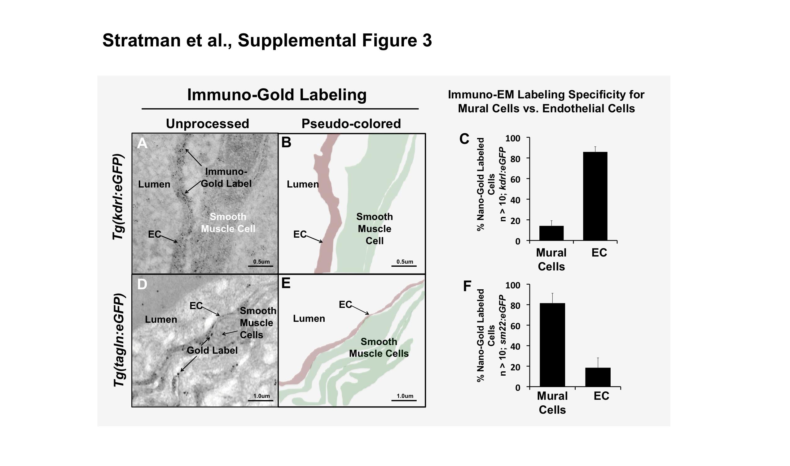
Immuno-EM confirms specificity of the tagln mural cell transgenic line. (A-C) Immuno-gold labeling of GFP of the Tg(kdrl:egfp) line shows specific labeling of the endothelium. (A) Low magnification and pseudo-colored interpretation of the endothelial (red) immuno-gold labeling. (B) Higher magnification of the endothelium shows more clearly the immuno-gold label. (C) Quantification of the number of immuno-gold labeled sites, showing that the labeling was largely endothelial specific. (D-F) Immuno-gold labeling of GFP of the Tg(tagln:egfp) line shows specific labeling of mural cells. (D) Low magnification and pseudo-colored interpretation of the mural cell (green) immuno-gold labeling. (E) Higher magnification of the mural cells shows more clearly the immuno-gold label outside of the endothelial cells lining the dorsal aorta. (F) Quantification of the number of immuno-gold labeled sites, showing that the labeling was largely mural cell specific. n = 10 image regions measured, regions collected from 2-3 individual fish. Scale bars = 0.5 or 1.0 μm as indicated. Mean ± s.e.m.