
Fig. 2
- ID
- ZDB-IMAGE-170203-2
- Publication
- Ling et al., 2017 - Distinct requirements of wls, wnt9a, wnt5b and gpc4 in regulating chondrocyte maturation and timing of endochondral ossification
- All Figures
- Figures for Ling et al., 2017
|
Fig. 2
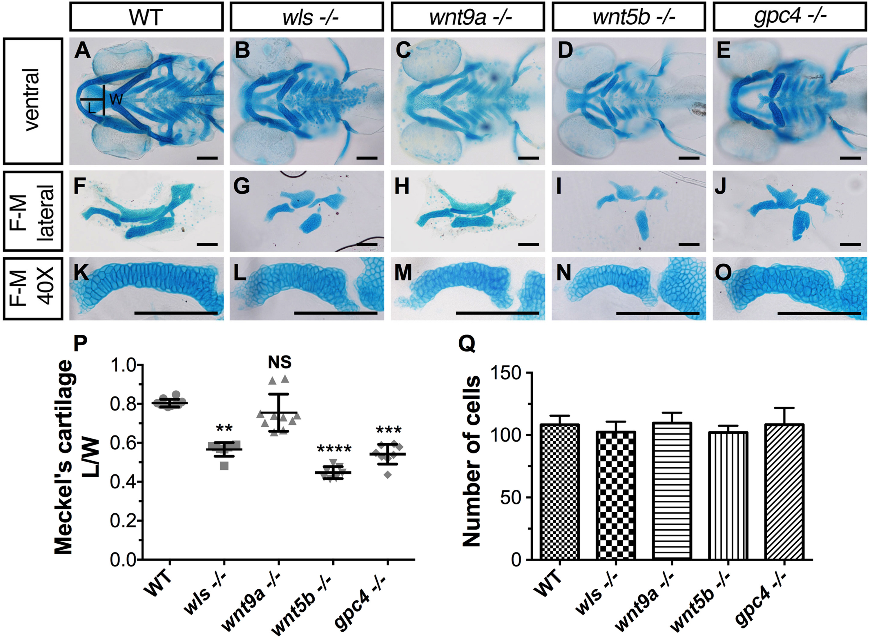
Cartilage morphogenetic defects inwls,wnt9a,wnt5bandgpc4mutants. (A-E) Ventral view of whole-mount 96 hpf Alcian blue stain of wls, wnt9a, wnt5b and gpc4. (F-J) Dissected flat-mounted lateral image of ventral cartilages. (K-O) 40X image of the Meckel's cartilage lateral view. Length (L) calculated from the midline tip of the Meckel's cartilage to the middle of the imaginary line (width (W)) between the retroarticular processes. (P) Meckel's cartilage length/width ratio measured from a clutch of imaged embryos (**p<0.01, ***p<0.001, ****p<0.0001; Kruskal-Wallis test with Dunn's multiple comparison test. Significance level at p<0.05). (Q) Number of cells from one-half of Meckel's cartilage was counted from figures K-O. No statistically significant difference in cell number across wls, wnt9a, wnt5b and gpc4 (p=0.1151, NS; One-way ANOVA). Scale =50 µm..
Reprinted from Developmental Biology, 421(2), Ling, I.T., Rochard, L., Liao, E.C., Distinct requirements of wls, wnt9a, wnt5b and gpc4 in regulating chondrocyte maturation and timing of endochondral ossification, 219-232, Copyright (2017) with permission from Elsevier. Full text @ Dev. Biol.