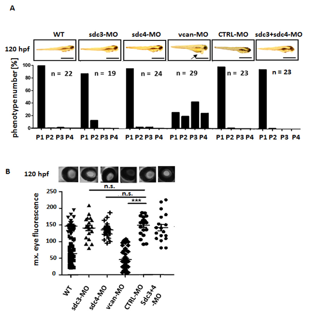
Fig. 4
Knockdown of syndecan 2 and versican causes edema and proteinuria in zebrafish larvae. (A) Edema quantification of zebrafish larvae at 120 hpf. Zebrafish were injected with a specific morpholinos for syndecan 3 (sdc3-MO, 100 μM), syndecan 4 (sdc4-MO, 100 μM), versican (vcan-MO, 100 μM), a scrambled control (CTRL-MO, 100 μM) or a combination of syndecan 3 (sdc3- MO, 100 μM) and syndecan 4 (sdc4-MO, 100 μM) at the one to four cell stage as indicated. The edema phenotypes of the larvae were categorized into 4 groups: P1 = no edema, P2 = mild edema, P3 = severe edema, P4 = very severe edema. (B) Corresponding fluorescent images of the retinal vessel plexus of Tg(lfabp: DBP:EGFP) of zebrafish larvae expressing a high molecular weight fluorescent protein in the circulation. Scatter graph presents maximum of circulating fluorescence intensity in the fish eye at 120 hpf, analyzed with image J. *** p<0.001, n.s. not significant; hpf: hours post fertilization.

