
Fig. S10
- ID
- ZDB-IMAGE-170110-24
- Publication
- Xiao et al., 2016 - Chromatin-remodelling factor Brg1 regulates myocardial proliferation and regeneration in zebrafish
- All Figures
- Figures for Xiao et al., 2016
|
Fig. S10
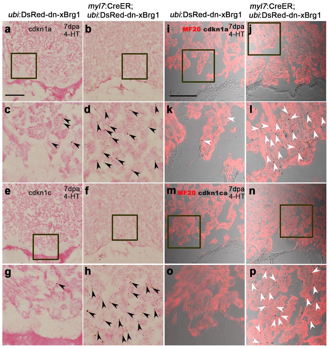
cdkn1a and cdkn1c are induced in Tg(myl7:CreER; ubi:DsRed-dn-xBrg1) transgenic hearts. (a-h) RNAScope in situ hybridization analysis with cdkn1a (a-d) and cdkn1c (e-h) probes on frozen sections of injured control Tg(ubi:DsRed-dn-xBrg1) hearts (a, c, e, g) and injured Tg(myl7:CreER; ubi:DsRed-dn-xBrg1) transgenic hearts (b, d, f, h) at 7dpa after 4-HT induction. Panels c, d, g and h are high-magnification images of areas in squares in panels a, b, e and f. Black arrowheads indicate the RNAScope signals. (i-p) Bright-field views of cdkn1a (i-l) and cdkn1ca (m-p) expression by RNAscope in situ hybridization, which were merged with MF20 antibody confocal images, on frozen sections of injured Tg(ubi:DsRed-dn-xBrg1) hearts (i, k, m, o) and injured Tg(myl7:CreER; ubi:DsRed-dn-xBrg1) transgenic hearts (j, l, n, p) at 7dpa after 4-HT induction. Panels k, l, o and p are high-magnification images of areas in squares in panels i, j, m and n. White arrowheads show RNAScope signals in cardiomyocytes. Scale bars, 100 μm.