
Fig. S7
- ID
- ZDB-IMAGE-170110-21
- Publication
- Xiao et al., 2016 - Chromatin-remodelling factor Brg1 regulates myocardial proliferation and regeneration in zebrafish
- All Figures
- Figures for Xiao et al., 2016
|
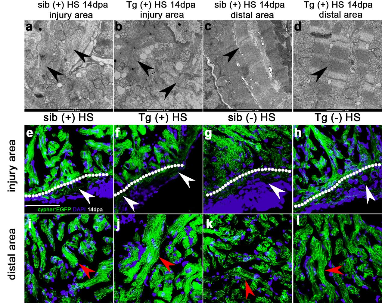
Fig. S7
Inhibition of Brg1 has no effect on cardiac sarcomere disassembly during heart regeneration. (a-d) Transmission electron microscopy images of myocytes of wt sibling (sib) (a, c), and Tg(hsp70:dn-xBrg1) transgenic (tg) heart (b, d) at 14 dpa. Note the normal sarcomere structures (black arrowhead) in distal cardiomyocytes from the injury site (c, d) and sarcomere disarray (black arrowhead) in cardiomyocytes in the injury site of both tg and wt hearts with heat shock treatment (a, b). Scale bars, 1μm. (e-h) Z-disks were labeled by using cypher-EGFP fusion protein. Cypher-EGFP-labeled sarcomere was all disarrayed in cardiomyocytes near the injury site (e-h), but was normal in distal area (i-l), of tg and wt sibling hearts at 14 dpa with or without heat-shock treatment. White arrowheads indicate sarcomere disarray (e-h) while red arrowheads indicate normal sarcomeres (i-l). DAPI co-stained for the nuclei; Scale bar, 50μm.